
Compilation of notes on embryology!
Contents:
- Notes on the Introduction to Embryology
- Notes on Ontogenetic Development and Embryology
- Notes on Descriptive and Comparative Embryology
- Notes on the Phases of Ontogenetic Development
- Notes on Experimental Embryology
- Notes on Blastogenesis and Embryogenesis
- Notes on Modern Embryology—Analytical Embryology
Note # 1. Introduction to Embryology:
Embryology is the study of structure and development of embryo, including the structure and development of male and female reproductive organs, fertilisation and similar other processes.
The Phanerogams (the flowering-plants) are also called spermatophytes (the seed bearing plants). These plants propagate mainly through seeds. The seed is a structure in which the embryo is enclosed. Adjacent to the embryo, foods are stored either inside the endosperm (albuminous) or in cotyledon (exalbuminous) for future use.
The Greek philosopher Aristotle (384-322 B.C.) is the ‘Father of the Study of Natural Sciences’. The greater part of his work is not available. With the available data, it seems that he did not recognise the presence of sex in plants. He believed that male and female plants were so blended that they generated on their own and the offspring developed from the superfluous (above what is enough) food of the plant.
Later Theophrastus (370-287 B.C), the ‘Father of Botany’ in third century B.C. referred the pollination of the date palm. After this, the nature of sexuality of plants was forgotten for about 2,000 years and his report was considered as useless. After a long gap, it was Nehemiah Grew who first clearly stated, in 1682, that the stamens are nothing but the male sex organs of a plant.
Later, in 1694, R. J. Camerarius suggested that the anthers are the male sex organs of a plant, while the ovaries with the styles were the female sex organs. After many years, J. G. Kolreuter (1761) fully confirmed the idea of Camerarius.
Later, Nawaschin (1898) had established the phenomenon of double fertilisation in Fritillaria tenella and Lilium martagon. Later on, the different aspects of embryology were studied by different workers. Most of the facts on development of gametophytes and embryo were discovered later.
During the 20th century, particular interest was developed on experimental embryology which includes the problems on storage and viability of pollen grains, effect of environmental factors on the growth of pollen tube, production of seedless fruits, embryo culture, induction of parthenogenesis etc. Thus embryology maintains an intimate association with genetics and physiology and opens many promising lines of research for the useful and economic development of crops.
Embryology played a significant role in consideration of systemic botany. Although Hofmeister and Strasburger pointed out the possibility of utilising embryological characteristics in taxonomy, it was Schnarf (1931) who first prominently brought into focus the role of embryology in taxonomy.
Note # 2. Ontogenetic Development as the Subject Matter of Embryology:
The aim of this article is to familiarize the reader with the basic facts and problems of the science of embryology. The name “embryology” is somewhat misleading. Literally it means the study of embryos. The term “embryo” denotes the juvenile stage of an animal while it is contained in the egg (within the egg envelopes) or in the maternal body.
A young animal, once it has hatched from the egg or has been born, ceases to be an embryo and would escape from the sphere pertaining to the science of embryology, if we were to keep strictly to the exact meaning of the word. Although birth or hatching from the egg is a very important occasion in the life of the animal, it must be admitted that the processes going on in the animal’s body may not be profoundly different before and after the hatching from an egg, especially in some lower animals.
It would be artificial to limit studies of the juvenile forms of animal life to the period before the animal is hatched from the egg or is born. It is customary, therefore, to study the life history of an animal as a whole and accordingly to interpret the scope of the science of embryology as the study of the development of animals.
The word “development” must be qualified in turn. In the sphere of biology with which we are concerned, the term “development” is used with two different meanings. It is used to denote the processes that are involved in the transformation of the fertilized egg, or some other rudiment derived from a parent organism, into a new adult individual.
The term development may, however, also be applied legitimately to the gradual historical transformation of the forms of life, starting with simple forms which might have been the first to appear and leading to the contemporary diversity of organic life on our planet. Development of the first type may be distinguished as individual development or ontogenetic development.
Development of the second type is the historical development of species or phylogenetic development. Phylogenetic development is often referred to as evolutionary development or simply evolution. Accordingly, we will define embryology as the study of the ontogenetic development of organisms. In this article we will be dealing only with the ontogenetic development of multicellular animals, the Metazoa.
In multicellular animals, the typical and most widespread form of ontogenetic development is the type occurring in sexual reproduction. In sexual reproduction new individuals are produced by special generative cells or gametes.
These cells differ essentially from other cells of the animal, in that they go through the process of maturation or meiosis, as a result of which they lose half of their chromosomes and become haploid, whereas all other cells of the parent individual, the somatic cells, are as a rule, diploid.
Once a cell has gone through the process of meiosis, it can no longer function as an integral part of the parent body but is sooner or later extruded to serve in the formation of a new individual. In multicellular animals there exist two types of sex cells – the female cells or ova, and the male cells or spermatozoa.
As a rule the two cells of the opposite sexes must unite in the process of fertilization before development can start. When the two gametes (the ovum and the spermatozoon) unite, they fuse into a single cell, the zygote, which again has a diploid number of chromosomes. The zygote, or fertilized ovum, then proceeds to develop into a new adult animal.
Side by side with sexual reproduction there exists in many species of animals a different mode of producing new generations—asexual reproduction. In asexual reproduction the off-spring are not derived from generative cells (gametes) but rather from parts of the parent’s body consisting of somatic cells.
The size of the part which is set aside as the rudiment of the new individual may be large or small, but in the Metazoa it always consists of more than one cell. The development of an animal by way of asexual reproduction obviously belongs in the same category as the development from an egg and should be treated as a special form of ontogenetic development.
To distinguish between the two forms of ontogenetic development, the term embryogenesis may be used to denote development from the egg, and the term blastogenesis may be used for the development of new individuals by means of asexual reproduction.
Note # 3. Descriptive and Comparative Embryology:
Although the correct understanding of ontogenetic development could be achieved only after the establishment of the cell theory, fragmentary information on the development of animals has been obtained since very ancient times. Aristotle had described the development of the chick in the egg as early as 340 B.C.
Many observations on development of various animals, especially of insects and vertebrates, were made in the seventeenth and eighteenth centuries. However, the data of embryology were first presented in a coherent form by Karl Ernst von Baer (1828). In his book “Ueber Entwicklungsgeschiechte der Tiere, Beobachtung and Reflexion,” Baer not only summed up the existing data and supplemented them by his original investigations but also made some important generalizations.
The most important of these is known as Baer’s law. The law can be formulated thus; “More general features that are common to all the members of a group of animals are, in the embryo, developed earlier than the more special features which distinguish the various members of the group.”
Thus the features that characterize all vertebrate animals (brain and spinal cord, axial skeleton in the form of a notochord, segmented muscles, aortic arches) are developed earlier than the features distinguishing the various classes of vertebrates (limbs in quadrupeds, hair in mammals, feathers in birds, etc.).
The characters distinguishing the families, genera, and species come last in the development of the individual. The early embryo thus has a structure common to all members of a large group of the animal kingdom and may be said to represent the basic plan of organization of that particular group. The groups having a common basic plan of organization are the phyla of the animal kingdom.
Baer’s law was formulated at a time when the theory of evolution was not recognized by the majority of biologists. It has been found, however, that the law can be reinterpreted in the light of the evolutionary theory. In its new form the law is known as the biogenetic law of Müller-Haeckel. Müller propounded the law in its new form and supported it by extensive observations on the development of crustaceans (1864). Haeckel (in 1868) gave it the name of the “biogenetic law” and contributed most to its wide application in biology.
According to Baer’s law, the common features of large groups of animals develop earliest during ontogeny. In the light of the evolutionary theory, however, these features are the ones that are inherited from the common ancestor of the animal group in question; therefore, they have an ancient origin.
The features that distinguish the various animals from one another are those that the animals have acquired later in the course of their evolution. Baer’s law states that these features in ontogeny develop at later stages. Briefly, the features of ancient origin develop early in ontogeny; features of newer origin develop late.
Hence, the ontogenetic development presents the various features of the animal’s organization in the same sequence as they evolved during the phylogenetic development. Ontogeny is a recapitulation of phylogeny. The repetition is obviously not a complete one, and the biogenetic law states that “ontogeny is a shortened and modified recapitulation of phylogeny.”
The shortening of the process is evident not only from the fact that what had once taken thousands of millions of years (phylogeny) is now performed in a matter of days and weeks (ontogeny), but also from the fact that many stages which occurred in the original phylogenetic development may be omitted in ontogeny.
The modifications arise mainly because the embryo at any given time is a living system which has to be in harmony with its surroundings if it is to stay alive. The embryo must be adapted to its surroundings, and these adaptations often necessitate the modification of inherited features of organization. A good example of such adaptation is the placenta in mammals.
The placenta is a structure developed by the embryo to establish a connection with the uterine wall of the mother and thus to provide for the nutrition of the embryo. This structure, though developed rather early in the life of the embryo, could not have existed in the adult mammalian ancestors. It is obviously an adaptation to the special conditions in which a mammalian embryo develops.
Even if the repetition of features of their ancestors in the ontogenetic development of contemporary animals is not complete, the fragmentary repetition of certain ancestral characters may still be very useful in elucidating the relationships of animals. As an example of this we may consider the formation of gill clefts or, at least, pharyngeal pouches in the ontogenetic development of all vertebrates.
In the aquatic vertebrates, such as Cyclostomata and fishes, the gill clefts serve as respiratory organs. In the adult state of terrestrial vertebrates, the pharyngeal pouches have disappeared completely or have been modified out of all recognition, and the function of respiration has been taken over by other organs—the lungs. Nevertheless, the pharyngeal pouches appear in the embryo.
In amphibians whose larvae are aquatic, the pharyngeal pouches at least temporarily serve for respiration. In reptiles, birds, and mammals, the pharyngeal pouches of the embryo do not serve for respiration at all. Their formation can be explained only as an indication that the terrestrial vertebrates have been derived from aquatic forms with functional gills. The paleontological evidence fully confirms this conclusion.
The systematic position of some animals cannot be recognized from adult structure, owing to profound modification acquired as a result of adaptation to very special conditions. Here the knowledge of the development sometimes throws unexpected light on true relationships. The adult ascidian is a sessile animal with no organs of locomotion and a nervous system of a very primitive nature.
The adult animal had been classed as a near relative of molluscs until Kowalevsky (1866) discovered that the larvae of the ascidians possess a well-developed dorsal brain and spinal cord, a definite notochord, and lateral bands of muscles (in short, organs that are typical for the vertebrates). The ascidians are therefore considered as belonging to the same phylum as the vertebrates, the phylum Chordata.
In the adult parasitic animal Sacculina, the organization of the animal is very much simplified in relation to the easy life that the parasite enjoys; it is reduced practically to a shapeless sack producing eggs and a system of branched rhizoids, by means of which the parasite is attached to its host, the crab, and absorbs the host’s body fluids on which it feeds.
It would be impossible to place Sacculina in any group of the animal kingdom if its development were not known. However, the larva of Sacculina is a typical arthropod, bearing a close similarity to the larvae of the lower crustaceans, the Entomostraca.
A rather similar larva is also found in the barnacles (Cirripedia) which, though possessing jointed legs like other arthropods, have lost the segmentation of the body in the adult state.
The attachment of the starfish larva, the brachiolaria, to the substrate while it is metamorphosing into the definitive form is an indication that the free-living echinoderms have been derived from sessile forms. This conclusion is again borne out by the evidence of paleontology.
Following the principles of Baer’s law and the biogenetic law, embryologists have systematically investigated the development of animals belonging to all the major groups of the animal kingdom. As a result of very extensive and painstaking investigations, a magnificent edifice of comparative embryology has been built.
Explaining Development – Theories of Preformation and Epigenesis:
Neither the description of morphological transformations occurring in the embryo nor the comparison of embryos and larvae among themselves and with the adult animals exhausts all the problems presented by the ontogenetic development of animals.
The fundamental problem presented by the existence of cyclical ontogenetic changes is the question – Why does ontogeny occur at all? What are the forces which produce the changes? How it is that, starting from a simple spherical cell, the process always ends in producing a highly complex and specific structure which, though varying in detail, reproduces with astonishing perseverance the same or almost the same adult form?
Attempts at solving this basic problem have been made ever since the human mind recognized the existence of development. For a long time the explanations proposed were purely speculative. Aristotle attempted to give a solution to the problem of ontogeny along the general lines of his philosophical teaching, distinguishing between the substance and the form of things.
The form appears in this concept as the creative principle. Aristotle further supposed that the substance for the development of a child is provided by the mother (in the form of nutrition) but that the creative principle is supplied by the father. He thus accounted also for the necessity of fertilization.
Although this treatment of the phenomena of development is completely contradictory to what we now know of the material basis of development (the parts played by the ovum and the spermatozoon), still the concept of a creative principle has turned up repeatedly in the teachings of embryologists up to the twentieth century.
In the seventeenth and eighteenth centuries, when all biological sciences developed rapidly, together with the physicochemical sciences, there existed a widespread theory explaining the ontogenetic development of animals. This was the theory of preformation. The theory of preformation claimed that if we see that something develops from the egg, and then this something must actually have been there all the time but in an invisible form.
It is common knowledge that in a bud of a tree the leaves, and sometimes also the flowers with all their parts, can be discovered long before the bud starts growing and spreading and thus exposing to view all that before was hidden inside, covered by the superficial scales of the bud. Furthermore, it was known that in a chrysalis of a butterfly the parts of the butterfly’s body—the legs, the wings, etc.—can be discovered if the cuticular coat of the chrysalis is carefully removed a few days before the butterfly emerges from the chrysalis.
Something of this sort was supposed to exist in the egg. All the parts of the future embryo were imagined to be already in the egg, but they were thought to be transparent, folded together, and very small, so that they could not be seen.
When the embryo began to develop, these parts supposedly started to grow, unfold, and stretch themselves, become denser and therefore more readily visible. The embryo, and therefore indirectly also the future animal, was preformed in the egg. Hence the theory is called the theory of preformation.
When spermatozoa were discovered in the seminal fluid, the relative significance of the ova and spermatozoa had to be accounted for. It is obvious that a preformed embryo cannot be present in both the egg and the spermatozoon. The preformationists were split therefore into two rival schools, the ovists and the animalculists. (The latter name comes from the word animalcule, as the spermatozoa were then called.)
The ovists asserted that the embryo was preformed in the egg. The spermatozoa then seemed superfluous, and in fact, they were declared parasites living in the spermatic fluid. On the other hand, the animalculists declared that the embryo was preformed in the spermatozoon and that the egg served only to supply nutrition for the developing embryo.
A lively discussion arose, which ended in favor of the ovists. The victory of the ovists was due to the discovery of parthenogenetic development in some insects, e.g., the aphids. If the egg could develop without fertilization, it was clear that the embryo could not be preformed in the spermatozoon.
The theory of preformation, although very popular in its time, did not satisfy all biologists, and opposing views, denying the existence of a preformed embryo in the egg, were proposed. The most important contribution in this field was the theory of epigenesis, proposed by Caspar Friedrich Wolff (1759).
In favor of his theory Wolff adduced his own observations on the formation of the chick embryo. In the earliest stages of the development of the chick he could not find any parts of the future embryo. Moreover, he found that the egg was by no means devoid of any visible structure; there was a structure present, but it was different from that of the later embryo.
Wolff found that the substance of which the embryo is composed is granular. Presumably the granules must have been the cells or their nuclei. These granules were later arranged into the layers which we now call the germinal layers. Wolff saw that by the formation of local thickenings in some parts of these layers, by thinning out in others, and by the formation of folds and pockets, the layers are transformed into the body of the embryo.
He concluded, therefore, that in the early egg there does not exist a preformed embryo but only the material of which the embryo is built. This material does not represent an embryo any more than a heap of bricks represents the house that will be built of them.
In both cases there had to be an architect who would use the material for a purpose that he had in mind. In the case of the developing embryo the architect was represented by a vital force, perhaps not essentially different from the “creative principle” postulated by Aristotle.
Note # 4. The Phases of Ontogenetic Development:
The processes leading to the development of a new individual really start before the fertilization of the egg, because the ripening of the egg and the formation of the spermatozoon, which constitute the phase of gametogenesis, create the conditions from which the subsequent embryogenesis takes its start.
In both oogenesis and spermatogenesis, meiosis, by discarding half of the chromosomes, singles out the set of genes which are to operate in the development of a particular individual. In both sexes the initial cells giving rise to the gametes are very similar and, as a rule, not essentially different from other cells of the body except that these cells are not involved in any of the differentiations serving to support the life of the parent individual.
In both sexes the first step in the production of gametes is a more or less rapid proliferation of cells by ordinary mitosis. The proliferating cells in the testes are known as the spermatogonia; the proliferating cells in the ovaries are called oogonia. Once proliferation ceases, the cells are called spermatocytes in the male and oocytes in the female. They then enter into a stage of growth and later into a stage of maturation.
Although the stage of proliferation is not essentially different in the male and female, the processes of growth and maturation in the two sexes differ to a very great extent. The cytoplasmic differentiations of the spermatozoon enable it to reach the egg by active movement and to fertilize it.
On the other hand, the egg cell accumulates in its cytoplasm substances which are used up during development—either directly, by becoming transformed into the various structures of which the embryo consists, or indirectly, as sources of energy for development. The elaboration in the egg cell of cytoplasmic substances to be used by the embryo and their placing in correct positions are essential parts of what occurs during the first phase of development.
The second phase of development is fertilization. Fertilization involves a number of rather independent biological and physiological processes. First, the spermatozoa must be brought into proximity with the eggs if fertilization is to occur. This involves adaptations on the part of the parents which insure that they meet during the breeding season, discharge their sex cells simultaneously in cases of external fertilization, or copulate in cases of internal fertilization.
Next the spermatozoa must find the egg and fuse with it. This egg-sperm fusion entails a very finely adjusted mechanism of morphological and physicochemical reactions. The egg is then activated by a spermatozoon and starts developing. A further rearrangement of the organ-forming substances in the egg is among the first changes that take place in the egg after fertilization.
The third phase of development is the period of cleavage. The fertilized egg is still a single cell, since the nucleus and cytoplasm of the spermatozoon fuse with the nucleus and cytoplasm of the egg. If a complex and multicellular organism is to develop from a single cell, the egg, the latter must give rise to a large number of cells.
This is achieved by a number of mitotic cell divisions following one another in quick succession. During this period the size of the embryo does not change, the cleavage cells or blastomeres becoming smaller and smaller with each division. No far-reaching changes can be discovered in the substance of the developing embryo during the period of cleavage, as if the preoccupation with the increase of cell numbers excludes the possibility of any other activity.
The whole process of cleavage is dominated by the cytoplasmic organoids of the cells, the centrosomes and achromatic figures. The nuclei multiply but do not interfere with the processes going on in the cytoplasm. The result of cleavage is sometimes a compact heap of cells, but usually the cells are arranged in a hollow spherical body, a blastula, with a layer of cells, the blastoderm, surrounding a cavity, the blastocoele.
The fourth phase of development that of gastrulation follows. During this phase the single layer of cells, the blastoderm, gives rise to two or more layers of cells known as the germinal layers. The germinal layers are complex rudiments from which are derived the various organs of the animal’s body.
In higher animals the body consists of several layers of tissues and organs, such as the skin, the subcutaneous connective tissue, the layer of muscles, the wall of the gut, and so on. All these tissues and organs may be traced back to three layers of cells—the aforementioned germinal layers. Of these the external one, the ectoderm, always gives rise to the skin epidermis and the nervous system.
The layer next to the first, the mesoderm, is the source of the muscles, the blood vascular system, the lining of the secondary body cavity (the coelom, in animals in which such a cavity is present), and the sex organs. In many animals, particularly the vertebrates, the excretory system and most of the internal skeleton are also derived from the mesoderm. The third and innermost germinal layer, the endoderm, forms the alimentary canal and the digestive glands.
The germinal layers are produced by the disappearance of a part of the blastoderm from the surface and its enclosure by the remainder of the blastoderm. The part that remains on the surface becomes ectoderm; the part disappearing into the interior becomes endoderm and mesoderm.
The disappearance of endoderm and mesoderm from the surface sometimes takes the form of a folding-in of part of the blastoderm, so that the simple spherical body becomes converted into a double-walled cup, as if one side of the wall of an elastic hollow ball had been pushed in by an external force.
This in-folding or pushing in of the endoderm and mesoderm is known as invagination, and the resulting embryo is known as a gastrula—whence the term gastrulation. The way in which the endoderm and mesoderm become separated from each other in the interior of the gastrula varies a great deal in different animals and cannot be described in this general review.
If the gastrula is formed by invagination, the cavity of the double-walled cup is called the archenteron, and the opening leading from this cavity to the exterior is called the blastopore. In animals in which the gastrula is formed in a different way—not by invagination—the cavity (archenteron) and the opening of the cavity to the exterior (blastopore) may still appear later on.
The archenteron, or part of it, eventually gives rise to the cavity of the alimentary system. The fate of the blastopore differs in the three main groups of Metazoa. In Coelenterata it becomes the oral opening. In Protostomia (including Annelida, Mollusca, Arthropoda, and allied groups) it becomes subdivided into two openings, one of which becomes the mouth and the other the anus. In Deuterostomia (including Echinodermata and Chordata) only the anal opening is connected in its development with the blastopore, the mouth being formed later on as an independent perforation of the body wall.
The whole of the lining of the alimentary canal does not always consist of endoderm; in all groups of animals the ectoderm may be invaginated secondarily at the oral or at both oral and anal openings to become a part of the alimentary canal. The parts of the alimentary canal lined by ectoderm are known as the stomodeum (adjoining the mouth) and proctodeum (adjoining the anus).
With the formation of the three germinal layers, the process of subdivision of the embryo into parts with specific destinies commences. This subdivision is continued in the next (fifth) phase of development, the phase of organogenesis (organ formation). The continuous masses of cells of the three germinal layers become split up into smaller groups of cells, each of which is destined to produce a certain organ or part of the animal.
Every organ begins its development as a group of cells segregated from the other cells of the embryo. This group of cells we will call the rudiment of the respective organ. The rudiments into which the germinal layers become subdivided are called primary organ rudiments.
Some of these are very complex, containing cells destined to produce a whole system of organs, such as the entire nervous system or the alimentary canal. These complex primary organ rudiments later become subdivided into secondary organ rudiments—the rudiments of the subordinated and simpler organs and parts.
The formation of the primary organ rudiments follows so closely on the processes of gastrulation that the two processes can hardly be considered separately. With the appearance of primary and secondary organ rudiments, the embryo begins to show some similarity to the adult animal or to the larva if the development includes a larval stage.
The sixth phase of development is the period of growth and histological differentiation. After the organ rudiments are formed they begin to grow and greatly increase their volume. In this way the animal gradually achieves the size of its parents. Sooner or later the cells in each rudiment become histologically differentiated; that is, they acquire the structure and physicochemical properties which enable them to perform their physiological functions.
When the cells in all the organs, or at least in the vitally important organs, have become capable of performing their physiological functions, the young animal can embark upon an independent existence—an existence in which it has to procure food from the surrounding environment.
In rather rare cases (in the nematodes, for instance) the young animal emerging from the egg is a miniature copy of the adult animal and differs from the latter only in size and the degree of differentiation of the sex organs. In this case the subsequent development consists only of growth and maturation of the gonads.
It is more usual, however, for animals emerging from eggs to differ from the adult to a greater extent; not only the gonads but also other organs may not be fully differentiated, or they may even be absent altogether and have to develop later. Sometimes the animal emerging from the egg possesses special organs which are absent in the adult but which are necessary for the special mode of existence of the young animal.
In this case the young animal is called a larva. The larva may lead a different mode of life from the adult, and therein lies one of the advantages of having a larval stage in development. The larva undergoes a process of metamorphosis when it is transformed into an animal similar to the adult.
The metamorphosis involves more or less drastic changes in the organization of the larva, depending on the degree of difference between the larva and the adult. During metamorphosis new organs may develop, so that morphogenetic processes become active again after a more or less prolonged period of larval life.
A secondary activation of morphogenetic processes may be produced in a different way. Many animals possess considerable plasticity and may be able to repair injuries sustained from the environment or caused experimentally. Lost parts may be regenerated, and this means that the developmental processes may sometimes be repeated in an adult or adolescent organism.
Asexual reproduction of animals involves the development of new parts and organs in animals that have already achieved the adult stage. All morphogenetic processes occurring in the later life of the animal, after the larval stage, or even when the adult stage has been achieved, will be dealt with as constituting a seventh and last phase of development.
Note # 5. Experimental Embryology:
Wolff’s observations, however, could not be considered as final in deciding between the alternative theories of preformation and epigenesis. In spite of what he actually observed, it was still conceivable that organs and parts of the body of the future embryo were represented in the egg by discrete particles, qualitatively different among themselves. The granules which he saw might have been different in their properties.
Even if the transformation of such qualitatively different parts into the organs of the embryo should have been more complicated than was envisaged by the crude preformistic theory, the principle of preformation might well have held true in spite of the apparent homogeneity of the material of which the embryo was supposedly made. Observation alone could not make further advances toward the solution of this problem, and further progress could be achieved only with the aid of experiment.
One of the experiments which are relevant to the preceding problem is the separation of the two cells into which the egg is divided at the beginning of development. If the theory of preformation is correct, we should expect that one of the two first blastomeres, containing one half of the egg material, should develop into an embryo lacking one half of its organs and parts.
If, on the other hand, the substances contained in the egg are but the building material used for the construction of the embryo, then it is conceivable that half of the material might be sufficient for making a complete embryo even if it may have to be on a diminished scale, just as the bricks prepared for the construction of a big house may be used for building two houses of a smaller size.
The first embryologist to see this way of solving the problem was Wilhelm Roux (1850-1924). Accordingly, he proceeded to test one of the first two cleavage cells in the common frog for its ability to develop. To achieve his end Roux destroyed one of the two cleavage cells with a red-hot needle (1888).
The embryo that was derived from the surviving cleavage cell was found to develop, at least at first, as if it were still forming a half of a complete embryo. In other words, the developing embryo was defective, as it should have been according to the theory of preformation.
It was found later, however, that the technique used by Roux was too crude. The damaged cleavage cell had not been removed, and it was the presence of this damaged cleavage cell, as was later found out, that caused the defects in the surviving embryo. If the two cleavage cells of the egg were separated completely, two whole and, except for their size, normal embryos could develop, one from each of the two cleavage cells.
This result was first found by H. Driesch (1891), working on sea urchin eggs, and later by Endres (1895) and Spemann (1901, 1903), working with eggs of newts. Eventually the experiment was repeated by Schmidt (1933) on the frog, the same animal that had served for the experiments of Roux. Schmidt found that if the two cleavage cells were completely separated, each could develop into a whole embryo.
The first experiments on the developing embryo were followed by many others, and soon a new science was born – experimental embryology.
Experimental embryology, in contrast to comparative embryology or descriptive embryology, uses experiment as a method of investigation. However, the use of the experimental method in itself does not create a science or a branch of science. New branches of science are created by novel viewpoints and novel problems set before science.
It was the problem of what ontogenetic development actually is, what the driving forces behind it are, that necessitated the application of experiment after the methods of speculation and of pure observation were found to be impotent in solving the problem.
Note # 6. Blastogenesis and Embryogenesis:
In the general survey of ontogenetic development, we have found it useful to consider what tasks have to be performed by the embryo before the final condition (the development of the new adult individual) is achieved. If from this same viewpoint we compare embryogenesis (development of the egg) with blastogenesis (development from a blastema in asexual reproduction), we see at once that the task is very much simpler in the latter case.
The process of producing a new individual is simplest in reproduction by fission, when the blastozooid is derived from half the parental organism and in this way is provided with a large proportion of the organs and parts which are necessary for making the new individual self-sufficient. What has to be done is the regeneration of missing parts.
The whole mechanism of regeneration, is brought into play, including the factors determining the regenerating parts. The remnant of the old individual determines the nature, position, and orientation of the newly differentiated organs. The polarity and bi-laterality of the parent organism prevail in the blastozooids.
The task of development is more complicated in the case of budding, since all organs and differentiated parts of the blastozooid have to be produced anew. Nevertheless, the initial system, the bud, always has a higher degree of complexity than a fertilized egg or even than a blastula as it occurs in embryogenesis. A typical bud, always consists of two layers of epithelial cells.
The young zooid is thus already in possession of the concentric stratification of body layers, a condition which in embryogenesis is achieved only after gastrulation. It is very note-worthy; however, that the layers formed in the blastozooid do not necessarily correspond to the germinal layers developing in embryogenesis.
In the case of budding in coelenterates, there is a closer correspondence between the outer and inner layers of the bud and the ectoderm and endoderm of the gastrula. The fate of the two layers is the same, but we have seen that the inner layer may be derived not from the endodermal epithelium of the parent but from a thickening in the ectodermal epithelium.
When we turn to the buds of tunicates, we find that the inner vesicle corresponds to the endoderm neither in its origin nor its fate. It has been shown that, although in some tunicates the inner vesicle may be derived from the endoderm of the parent (in the form of an outgrowth from the pharyngeal epithelium), it can also be derived from mesoderm (epicardium, mesodermal septum of the stolon, blood cells) or even ectoderm (lining of the atrial cavity).
What is even more important is that the inner vesicle gives rise to parts derived in embryogenesis from any of the three germinal layers. In spite of the diversity of origin of the inner vesicle, its later differentiation shows a considerable degree of uniformity in different tunicates.
After the bud has grown to a certain degree—a minimal size, varying of course in different species, is essential — it becomes constricted at least partially from the parent zooid. Then folds start sub-dividing the inner vesicle into sections. (The following description refers to the development of the blastozooid in Botryllus.)
First, two folds cut in from what will be the distal part of the new zooid, subdividing the inner vesicle into a median part, which will become the branchial chamber, and two lateral parts, which give rise to the atrial cavity (Fig. 507). (In embryogenesis, the atrial cavity develops as a pair of invaginations of the ectoderm which partially fuse and later have a common opening to the exterior.)
In addition to these three main sub-divisions, further smaller pocket-like evaginations of the vesicle appear. One evagination near the anterior end of the central cavity, gives rise to the nerve center. (In embryrogenesis, the nerve ganglion is a remnant of the neural tube formed by infolding of the ectodermal neural plate, as in Amphioxus and in vertebrates.) Two pockets at the posterior end give rise to the intestinal loop (including the esophagus and the stomach) and to the pericardium, respectively.
(In embryogenesis the intestine is, of course, of endodermal origin, and the pericardium develops from mesodermal mesenchyme.) Because the initial state of the system in blastogenesis is not the same as that in embryogenesis, the course of development is different. The morphogenetic processes in budding appear to be simpler and more straightforward than in embryogenesis.
The actively developing part, which is the inner vesicle (the epidermis of the bud is a differentiated tissue all the time, specialized in secreting the cellulose mantle), proceeds directly to the formation of organ rudiments, omitting the stage of germ layer development. In one further respect the morphogenetic processes in budding are simpler than in embryogenesis: The new individual inherits its polarity directly from the parent zooid.
The point of attachment of the bud to the maternal body or the stolon always becomes the proximal end of the blastozooid. In the development of gemmules, the task of producing a new individual becomes most complicated and approaches that of the development of the egg.
The special difficulties encountered are:
1. The germination of the gemmule occurs after the death and decomposition of the parent animal; consequently, the polarity of the new individual has to be worked out by itself, and the parent body is no longer there to influence the polarity of the offspring.
2. The complete homogeneity of the contents of the gemmule (in the case of the gemmules of sponges) deprives the new individual of any remnant of morphological organization. The structure of the new sponge has to be established by the interaction of practically independent cells.
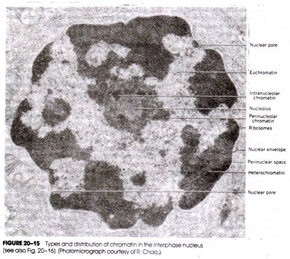
The interior of the gemmule consists of only one type of cell, the archeocytes, which are rather large, cells containing platelets of glycoprotein. Even before the germination of the gemmule, some of the archeocytes become activated; they start dividing and in so doing give rise to smaller and smaller cells, very much like the blastomeres which diminish in size as cleavage progresses. The glycoprotein platelets gradually disappear; the nuclei become richer in chromatin, a usual characteristic in actively growing and metabolizing cells.
These small and active cells have been referred to as histioblasts—cells producing tissues. When the gemmule germinates, the contents of the gemmule crawl out through the micropyle and form an irregular mass, surrounding the empty shell of the gemmule (Fig. 508a). Both the histioblasts and the remaining glycoprotein-containing archeocytes leave the shell. Outside the shell, the division of archeocytes and their conversion into histioblasts continue (Fig. 509).
The histioblasts now become arranged into an irregular meshwork (Fig. 508b), cavities appear, and some of the histioblasts surrounding these cavities differentiate into choanocytes. Other histioblasts become epidermal cells, scleroblasts, pore cells, or mesenchymal cells arranged in a typical way, so that the mass of cells soon becomes a small sponge, with a system of internal canals with ciliated chambers, ostia, etc.
It may be significant that the sponges are capable of reconstituting their structure after complete dis-aggregation. The development of the gemmule proceeds along very much the same lines; the development is direct in the extreme, the individual cells differentiating and taking up their positions in the whole.
A review of different forms of asexual reproduction reveals a principle of general significance. It shows that the complexity of morphogenetic processes is largely determined by the degree of difference that exists between the initial stage of development and the final condition.
The initial stage in embryogenesis is a single cell; therefore a period of cleavage is necessary, which brings the system into a multicellular condition. In asexual reproduction, the initial system is already multicellular, and cleavage falls away.
(Something resembling cleavage occurs, in the gemmules of sponges, where the archeocytes accumulate food reserves, similarly to oocytes in the ovary.) In asexual reproduction the initial system may have the cells arranged in more than one layer, and this makes gastrulation dispensable. What remains of the main periods of development are organogenesis, differentiation, and growth.
A second very suggestive fact which emerges from a study of asexual reproduction is that, given a normal environment, the organization of the animal’s body is entirely determined, in the last instance, by the hereditary constitution of the species-specific cells. The structure of the egg cell with its polarity and the heterogeneous arrangement of cytoplasmic substances is a mechanism which provides for the orderly course of differentiation of the cells in a developing embryo.
This mechanism is, however, dispensable; the same egg product can be attained starting from a different initial constellation, provided that the cells have the same hereditary constitution. In principle, the orderly organization of an animal’s body should be attainable by the interaction of an assortment of different types of cells produced on the basis of species-specific hereditary potentialities. In practice, this is possible only in relatively very simple biological systems.
Note # 7. Modern Embryology—Analytical Embryology:
After the middle of the present century embryology had got caught up in the new trend that developed in biological science. Early in the century, the background for this new trend was established mainly by the work of T. H. Morgan and his school.
This work proved that the units of heredity, the genes, are arranged in linear order in the chromosomes of the cells. Analysis shows that chromosomes consist of several chemical components- deoxyribonucleic acid (DNA), ribonucleic acid (RNA), and proteins.
In an epoch-making paper published in 1953, Watson and Crick suggested that the deoxyribonucleic acid, as found in the chromosomes, consists of pairs of very elongated molecules twisted spirally around each other in a double helix. Each strand of the helix is made up of a number of units, the mononucleotides, which differ from one another only in the nitrogenous base (i.e., adenine, thymine, guanine, or cytosine) which each contains.
The bases form two pairs, which structurally “fit” together, so that in the intertwined double helix adenine always links with thymine, and guanine with cytosine. Further work made it clear that the arrangement of the bases in the molecule of deoxyribonucleic acid contains a code for the proteins that may be synthesized by a particular species of organism.
The code is essentially a series of “triplets”—groups of three bases which correspond to one amino acid in a polypeptide (protein) chain. Thus, a sequence of triplets in the DNA determines a sequence of amino acids in a protein molecule, and the section of the deoxyribonucleic acid molecule containing this sequence is the essential part of what geneticists call a “gene.”
The “genetic code,” showing which sequences of bases correspond to which of the 20 amino acids constituting most of the proteins in the organic world. Note that several different triplets in the DNA may code for the same amino acid.
Between the genetic code in the chromosomal DNA and the cell proteins, there are certain intermediate steps. The “message” contained in the DNA must first be copied in the form of a ribonucleic acid molecule, whose nucleotide sequence is complementary to the nucleotide sequence of the DNA (except that uridine takes the place of thymine). This is the “transcription” phase. The code is then contained in an RNA molecule (“messenger” RNA).
Two further kinds of ribonucleic acid are modeled on the DNA- the ribosomal RNA, which together with certain proteins forms small (± 200 Å in diameter) particles, the ribosomes; and the transfer RNA, which is involved in bringing the correct amino acid to the ribosome, where the amino acids become arranged and joined together in the correct sequence according to the code contained in the messenger RNA. This procedure constitutes the “translation” phase.
The importance of these discoveries for embryology derives from the following considerations. It has become evident that all the properties of any organism are determined in the last instance by the sequence of base triplets in the DNA molecules. Furthermore, it is accepted that the sequence of the base triplets directly determines what kinds of proteins can be produced by an organism.
All other manifestations specific to any organism, whether morphological or physiological, depend more or less directly on the assortment of proteins coded for by the hereditary DNA. This new way of looking at the organic world shifts the problem of ontogenetic development directly into the realm of molecular relationships. It also makes possible, in principle, the construction of a complete theory of development.
Such a theory would start with the triplet sequences in the DNA and would show first how these sequences are “read out” by transforming them into an array of proteins, placed and distributed in an organized way in space and time, and then would show how the proteins, acting partly on their own and partly through other chemical components, produce the complicated system that is an adult organism.
A whole array of new techniques has been mobilized in working toward such a theory of development. Electron microscopy has made great advances after the mid 1950’s, when methods were developed for embedding tissues in plastics and for cutting ultrathin sections for the study of the fine structure of cells. Refined methods of chemical analysis, such as chromatography, electrophoresis, ultracentrifugation, and the use of radioactive tracers, have been put at the disposal of embryologists.
With the change in the theoretical background and techniques, a subtle change has permeated the work of embryologists. The aim of investigation is no longer the study of the development of any particular animal, or any group of animals, but the discovery of the basic principles and processes of development. This trend in science may perhaps be called analytical embryology, and this is what “modern” embryology actually is.
It must be realized that analytical embryology can proceed only on the basis of knowledge provided by descriptive embryology, because after all, it is the actual course of the transformations that has to be explained by the theory of development, of comparative embryology, because it is necessary to know of how general a significance any particular phenomenon of development is, and of experimental embryology, because it has revealed the causal relationships of many developmental processes.