In this article we will discuss about:- 1. Meaning of Cleavage 2. Planes of Cleavage 3. Types 4. Effects of Yolk 5. Mechanism 6. Chemical Changes 7. Different Chordates 8. Importance in Embryonic Pattern.
Contents:
- Meaning of Cleavage
- Planes of Cleavage
- Types of Cleavage
- Effects of Yolk in Cleavage
- Mechanism of Cleavage
- Chemical Changes during Cleavage
- Cleavage in Different Chordates
- Importance of Cleavage in Embryonic Pattern
1. Meaning of Cleavage:
Fertilization results into the formation of zygote. The process of segmentation (cleavage) immediately follows fertilization or any other process which activates the egg. Cleavage consists of division of the zygote into a large number of cellular entities. The cells which are produced during segmentation are called blastomeres.
At first, the cells remain closely associated, but later on they form the lining of a hollow sphere called blastula. The blastula contains a cavity named blastocoel and its outer covering is designated as blastoderm. The formation of blastula culminates the cleavage period.
The process of segmentation prepares the groundwork for the future design of the embryo by producing adequate number of cells. The cleavage also establishes the fundamental conditions for the initiation of next developmental stage —Gastrulation.
2. Planes of Cleavage:
During early cleavage, distinct geometrical relationships exist between the blastomeres, i.e., each plane of cell-division bears a definite relationship with each other.
The planes of division are:
a. Meridional plane of cleavage:
When a furrow bisect both the poles of the egg passing through the median axis or centre of egg it is called meridional plane of cleavage. The median axis runs between the centre of animal pole and vegetal pole.
b. Vertical plane of cleavage:
When a furrow passes in any direction (does not pass through the median axis) from the animal pole towards the opposite pole.
c. Equatorial plane of cleavage:
This type of cleavage plane divides the egg halfway between the animal and vegetal poles and the line of division runs at right angle to the median axis.
d. Latitudinal plane of cleavage:
This is almost similar to the equatorial plane of cleavage, but the furrow runs through the cytoplasm on either side of the equatorial plane.
3. Types of Cleavage:
Considerable amount of reorganisation occurs during the period of cleavage and the types of cleavage depend largely upon the cytoplasmic contents.
Different types of cleavage encountered in different eggs are catalogued below:
a. Holoblastic op total cleavage:
When the cleavage furrows divide the entire egg.
It may be:
Equal:
When the cleavage furrow cuts the egg into two equal cells. It may be radially symmetrical, bilaterally, symmetrical, spirally symmetrical or irregular.
Unequal:
When the resultant blastomeres become unequal ir size.
b. Meroblastic cleavage:
When segmentation takes place only in a small portion of the egg resulting in the formation of blastoderm, it is called meroblastic cleavage. Usually the blastoderm is present in the animal pole and the vegetal pole becomes laden with yolk which remains in an tihcleaved state, i.e., the plane of division does not reach the periphery of blastoderm or blastodisc.
c. Transitional cleavage:
In many eggs, the cleavage is atypical which is neither typically holoblastic nor meroblastic, but assumes a transitional stage between the two.
4. Effects of Yolk in Cleavage:
The fertilized egg in most cases contains yolk, which are inert bodies. During division these bodies exert mechanical influences. In the egg of Amphioxus, the yolk is thin and remains uniformly distributed. Therefore the division is complete and early divisions occur at a very quicker rate.
The amphibian egg contains yolk which is localised at the vegetal pole. Here division initiates from the animal pole and extends towards the vegetal pole, where the progress of cleavage slows down considerably.
Consequently, the animal pole divides faster than the vegetal pole. The eggs of reptiles and birds are fully laden with large masses of yolk, thus restricting the cytoplasm and nucleus on the periphery as a circular disc on the animal pole. Here the lines of cleavage divide only the small animal pole region. Such effects of yolk on cleavage pattern influence the pattern of further development.
5. Mechanism of Cleavage:
The incidence of cleavage provides unique opportunity to study the mechanism of cell division and specially the role of different cell organelles during division.
Opinions differ regarding the accumulation of force for the initiation of cleavage and following factors are believed to be responsible for controlling the cleavages:
(a) Localised expansion of cortex.
(b) Increased stiffness of the cortical cytoplasm.
(c) Increase of tangential force activity in the cortex.
(d) Contractile nature of the regions near the cortex and
(e) Formation of new cell membrane from the subcortical cytoplasm.
Though the abovementioned factors are not clearly understood, it is evident that three structures present within the cell: Cortical layer, Spindle structures and Chromosomes play the important part.
The energy which is required during the process is supplied by the metabolic activity of the developing egg. Besides the factors involved in segmentation, there are cleavage laws which govern the behaviour of the cells during cleavage.
Sach’s rules:
The blastomeres tend to divide into identical daughter cells and a cleavage furrow tends to cut the previous cell at right angles.
Hart wig’s laws:
The position of nucleus is vital and it tends to lie at the centre of the protoplasmic content of the cell. The nucleus exerts influence on cleavage. The long axis of mitotic spindle usually coincides with the long axis of the protoplasmic content. During cleavage the long axis of the protoplasm has the tendency to cut transversely.
Balfour’s law:
The rate of cleavage is inversely proportional to the amount of yolk material present in the egg.
6. Chemical Changes during Cleavage:
Significant chemical changes go on in the fertilized egg during cleavage.
They are:
Increase of nuclear material:
During cleavage a steady increase in nuclear material (predominantly DNA) is observed. Cytoplasm of the egg is the source of such nuclear material. Cytoplasmic DNA contained in mitochondria and yolk platelets are available.
RNA synthesis:
During cleavage messenger RNA (mRNA) and transfer RNA (tRNA) are synthesised during cleavage, especially in late stages.
Synthesis of proteins:
Throughout the period of cleavage there is steady and spectacular increase in protein synthesis.
7. Cleavage in Different Chordates:
The pattern of cleavage differs in different animals. The following account will give an idea of the process of cleavage in different chordates.
a. Amphioxus:
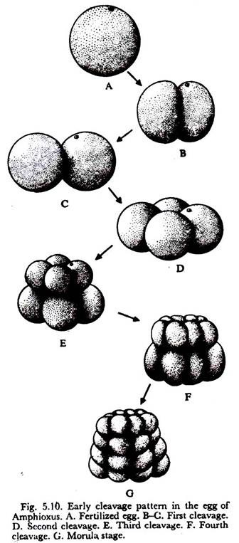
The cleavage in Amphioxus is typically holoblastic (Fig. 5.10). The first cleavage is meridional. The second cleavage is also meridional but at right angle to the first one. Four equal blastomeres are produced. The third cleavage is latitudinal and occurs slightly above the equatorial plane resulting in the production of eight blastomeres—four are smaller called the micromeres and four are larger known as the macromeres.
The micromeres are situated towards the animal pole and the macromeres towards the vegetal pole. The fourth cleavage is meridional which involves all the eight cells resulting in the formation of eight micromeres and eight macromeres. The fifth cleavage planes are latitudinal.
Each micromere is divided into an upper and lower micromere and each macromere likewise divides to form an upper and lower macromere. The fifth cleavage planes produce thirty-two blastomeres. The sixth cleavage planes are nearly meridional involving all the thirty-two cells resulting in sixty-four cells.
At the 64-cell stage a conspicuous space is produced at the centre and this space becomes filled with a fluid. When the eighth cleavage planes take place, the blastula becomes pear- shaped and the blastocoel becomes large.
b. Frog:
The egg of frog is telolecithal with a considerable amount of yolk localized towards the vegetal pole. The cleavage is holoblastic in nature, but differs considerably from that of Amphioxus because of larger quantity of yolk.
The first cleavage plane is meridional which occurs at about 3-3½ hours after fertilization. But the time depends largely on extrinsic factors. The first cleavage starts at the animal pole and gradually travels towards the vegetal pole. Thus the egg is bisected along the poles. Two blastomeres of equal size are produced. The second cleavage is almost meridional but oriented at right angles to the first cleavage plane (Fig. 5.11).
The four blastomeres thus produced are not qualitatively identical, because the grey crescent material is present in two of the four blastomeres. Each blastomere contains dark pigment at the animal pole and yellowish yolk towards the vegetal pole. The third cleavage is latitudinal and occurs at right angles to previous cleavage planes but passes slightly above the equator.
The furrow produces eight unequal blastomeres, four micromeres in the animal hemisphere and four macromeres in the vegetal part. The fourth cleavage planes are meridional which involve the micromeres first and pass on slowly towards the yolk-laden macromeres of the vegetal pole.
In Amphioxus, the cleavages occur in a synchronous fashion, while in frog considerable degree of irregularities (asynchronism) appear in later stages. But it is certain that the micromeres always continue to divide at a faster rate than do the macromeres.
At the eight-celled stage, a small space makes its appearance between the four micromeres. As development goes on, this space becomes conspicuous and forms the blastocoel. The floor of the blastocoel is formed of macromeres. The blastocoel (or segmentation cavity) is eccentrically located and becomes displaced towards the animal pole as development proceeds.
c. Chick:
Typical meroblastic cleavage occurs in chick, where the segmentation activity is restricted only at the blastodisc or germinal disc (Fig. 5.12). Thus the cleavage is incomplete.
The first cleavage starts as a meridional furrow near the centre of the blastodisc at about 4½ hours after fertilization when the egg reaches the isthmus of oviduct. This furrow cuts across the blastodisc and passes towards the vegetal pole but does not reach the pole. The second cleavage is also meridional, but approximately at right angles to the first one. The third cleavage is vertical.
The fourth cleavage is also vertical but the division is not synchronous. As a consequence eight central cells encircled by twelve marginal cells are produced. From this point onward the cleavage becomes irregular and a disc containing smaller cells appears.
This disc remains firmly connected with the underlying yolk. Soon a cleft appears which separates the disc in the middle from the underlying yolk. The new cavity in between is known as sub-germinal space (Fig. 5.13).
Thus at the end of segmentation, the disc contains many-layered small cells which are connected with the yolk only at the periphery. This disc is then termed as blastoderm, the cells of which still continue to divide.
The peripheral part which lies in contact with yolk possesses granular cells called area apaca and the inner layer having clear portion is called area pellucida. At one end of area opaca, aggregation of cells takes place. This denotes the formation of future posterior side.
d. Rabbit:
The egg of rabbit is small and does not contain any yolk (i.e. alecithal type of egg). The cleavage is holoblastic and nearly equal. Irregularities and a synchronism become the rule in the cleavage of rabbit like all other eutherian mammals.
The first cleavage is vertical resulting in the formation of two unequal blastomeres. The second cleavage is also vertical but runs at right angle to the first. The third cleavage is horizontal but slightly above the equator.
Subsequent divisions are rapid and irregular. The blastomeres thus produced become clustered together to form a solid cellular ball called morula. Two types of cells (small and large) are recognised in the morula.
The large cells lie at the centre. Soon a cavity appears inside the cell mass on one side. The cavity gradually increases which shifts the central cell mass to one side. The stage is called blastocyst stage. The inner cell mass in the centre is attached with the outer cell layer (trophoblast) of the blastocyst.
The cavity is called the blastocoel or sub-germinal cavity (Fig. 5.13A) which is filled with a fluid. The inner cell mass remains attached at the embryonic knob towards the animal pole. From this embryonic knob, the embryo arises. The trophoblast which encloses the blastocoel and the embryonic knob participates in the formation of placenta. The trophoblastic cells overlying the embryonic knob is called cells of Rauber.
8. Importance of Cleavage in Embryonic Pattern:
The cleavage phase of development and blastulation are extremely significant, because the blastoderm is morphologically elaborated in such a way that the important presumptive organ forming areas of the future embryo are segregated into definite districts of the blastoderm.
Such orientation of the organ forming areas in the blastoderm permits an ordered movement of these areas during gastrulation to take up their fateful position. So the period of cleavage and blastulation is regarded as the phase of preparation for future differentiation.
The cells which are produced at the end of segmentation resemble the zygote—but do they possess the same potentiality as the zygote itself. Driesch (1891), in order to get an answer, separated the two blastomeres at the two-celled stage and found that both the blastomeres developed into complete embryos.
His conclusion was that each blastomere has the full potentiality to be an entire embryo. But in 1900, Roux showed that if one of the blastomeres of the two-celled stage is killed, the remaining one produces ‘half embryo’.
He claimed that each cleavage results into the segregation of specialization in the blastomeres and this is irreversible. This experiment demonstrates that an organising or controlling centre is elaborated to control the development process.
The experiment of Spemann and others have shown that it is the grey crescent region which plays the vital role in the process of determination and the blastomeres which are formed due to segmentation are neither completely regulative nor irreversibly determined.
Fig. 5.14 shows the importance of grey crescent in the development of amphibian embryo. It has been experimentally established that the grey crescent in the amphibian blastula transforms into the dorsal lip of the blastopore which acts as an instigator and controller of the gastrulation process.


