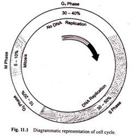
The following points highlight the four major phases of the cell cycle. The phases are: 1. G1 (gap1) phase 2. S (synthesis) phase 3. G2 (gap 2) phase 4. M (mitosis) phase.
Cell Cycle: Phase # 1. G1 Phase:
The G1 phase is set in immediately after the cell division. It is characterised by a change in the chromosome from the condensed mitotic state to the more extended interphase state and by a series of metabolic events leading to initiation of DNA replication. G, phase of cell cycle varies in length from cell to cell within the same cell population.
The length of this phase is more than the other three phases. This period represents in general 25 to 40% of the generation time of a cell. The cause of variability in G1 is not known, although there is an evidence to suggest, that the protein content of the cell possibly determines the length of G1.
The G1 is a very significant phase of cell cycle in the sense that the cells which stop dividing normally remain arrested in this phase. Within G1 phase, there is a definite check point at which DNA synthesis is initiated and once the biochemical events associated with that point have occurred the cell proceeds towards division.
There are specific molecular signals at that point which decide whether a cell has to continue its proliferation or it has to specialize into a permanent or non-dividing cell.
During G1, phase the chromatin fibres become slender, less coiled and fully extended and more active for transcription. The nucleus appears under light microscope to contain a network of delicate fibres. The transcriptional activities result in the synthesis of rRNA, tRNA, mRNA and a series of molecules required for the initiation of DNA synthesis.
The events which lead to the initiation of DNA synthesis include synthesis of enzymes and other proteins required for DNA synthesis.
Cell Cycle: Phase # 2. S Phase:
After G1, phase there comes the S phase. Biochemically, it is a phase of active DNA and histone synthesis. During S period doubling of the slender fully extended chromosomes takes place which is accomplished by doubling of DNA and the associated proteins in the chromosomes.
The replication of DNA is semiconservative and discontinuous. J.H. Taylor in 1957 labelled the root tip cells of broad bean with thymidine labelled with tritium (3H ).
The labelled cells were found to contain labelled chromatin during S period in auto-radiographs. Such labelled cells were allowed to go through one more division cycle in absence of label and then it was found that at any point along each chromosome, only one chromatid was labelled.
This indicated semiconservative replication of DNA. In bacteria there is a single replication point and replication of DNA proceeds bidirectionally from that point.
In eukaryotes, each chromosome has a number of replication units (RUs) or replicons, each of which has a specific origin and two termini for replication process. Electron microscopy and autoradiographic studies have indicated that the replication of DNA begins by the opening of a “bubble” in DNA resulting into two forks.
These forks migrate bidirectionally as the DNA is synthesised until they reach termination points. There are specific nucleotide sequences which mark the initiation and termination points. Careful examination of electron micrographs of replicating chromosomes shows the spacing between the replication units which, in a wide range of eukaryotes, ranges from 7 to 100 mm (30,000 to 300,000 base pairs).
After the termination process, the adjacent replication forks 166 Cytology are fused. Since DN A replication is dependent on protein and RNA synthesis for the overall replication of chromosome, it is necessary that new proteins must be synthesised.
Although some of the histone protein is synthesised during G1, phase, most of it is synthesised during S phase. Further, synthesis of ribosomal RNA must continue from G1, to S phase if DNA synthesis is to start.
The eukaryotic chromosomes consist of DNA-histones complexes, called nucleosomes. When the DNA is replicated and new histones are synthesised the two are complexed to form nucleosomes.
The distribution of old and newly synthesised histones in the nucleosomes of newly replicated DNA is not clearly understood but it is clear that the old octamers of histones are conserved from generation to generation and the octamers of newly synthesised histones are used at the replication forks to form new nucleosomes.
Cell Cycle: Phase # 3. G2 Phase:
G2 phase follows the S phase. The metabolic significance of this phase is not fully understood as yet. This phase represents to some 10 to 25% of the generation time of the cell. In G2, the chromosome consists of two strands or chromatids. The cells that are arrested at the transition between S and G2 will have twice the usual amount of DNA. Tetraploid nuclei are occasionally found in various tissues, as for example, in liver cells, heart muscles etc.
The critical event at G2, check point is concerned with the condensation of chromatin. The condensation process involves high order folding of the chromatin fibre but the mechanism for this is poorly understood. The chromatin condensation is associated with accumulation of a cytoplasmic substance.
The existence of chromatin condensing factor has been demonstrated by R.T. Johnson and P.N. Rao (1970, 74) by using Sendai virus to fuse He La cells (a type of cell derived from human cancer cell) at different stages of cell cycle. When a mitotic cell was fused with a cell at G2 phase, chromatin condensation took place at once in G2 nucleus, producing normal looking chromosomes.
When a mitotic cell was fused with a cell at G1, phase chromatin condensation was demonstrated in G1, nucleus also. Similarly, condensation of chromatin in S nucleus was also observed when mitotic cell was fused with a cell at S phase, although multiple fragments of condensed chromatin instead of complete chromosomes were seen.
The chromatin condensation is thought to be associated with, and perhaps due to, phosphorylation of histone H.
The syntheses of RNAs and proteins occur during most of the cell cycle, but the two processes may be inhibited during S phase and almost completely suppressed during late prophase. Another important event that takes place during G2 period is the synthesis of the proteins tubulin which is later polymerized to produce microtubules that make up spindle apparatus during M phase.
Cell Cycle: Phase # 4. M Phase:
M phase follows G2 phase. During this phase the cell divides into two daughter cells. The chromosomes are duplicated during interphase and they are distributed to the progeny cells by division process. After M phase, the resulting daughter cells then enter the G1, phase of next cell cycle. The cells which after completion of mitosis do not enter G1, are referred to as G0 cells.
During M phase, RNA synthesis stops at late prophase and resumes at telophase. The protein synthesis drops drastically and during that period RNA synthesis also stops. These changes in the synthetic activity may be attributed to the non-availability of transcription sites owing to highly condensed state of the chromosomes. If no mRNA is available to ribosomes, protein synthesis cannot take place.
The nucleolus and nuclear wall break down goes side by side with the inhibition of RNA and protein synthesis. These structures are reformed in daughter nuclei in late telophase when the synthesis of RNA and proteins resume. The molecular signals for these changes are not known. The behaviour of chromosomes during different phases of cell cycle, as illustrated by De Robertis et al. (1975) is shown in (Fig. 11.1).
