In this article we will learn about Chlamydomonas. After reading this article we will learn about: 1. Systematic Position of Chlamydomonas 2. Occurrence of Chlamydomonas 3.Structure 4. Reproduction.
Contents
Systematic Position of Chlamydomonas:
Occurrence of Chlamydomonas:
Chlamydomonas is a large genus and is found almost in all places. It is represented by about 400 species (Prescott, 1969). Chlamydomonas is simple, unicellular, motile fresh water algae. It is mainly found in fresh water rich in nitrogen salts and organic matter. It is also found in stagnant water of ponds, pools, ditches, water tanks, sewage tanks and in slow running water.
Chlamydomonas is planktonic algae and makes surface of water appear green. Some species of Chlamydomonas are terrestrial, they grow on moist soil surface, in rice fields and on banks of rivers and lakes. Palmella stages of genus make scum on soil surfaces. Some species are found in salty brackish water e.g., C. halophila, C. ehrenbergii.
Chlamydomonas is also found as cryophytes i.e., growing on snow e.g., C. nivalis causes red snow due to presence of red pigment haematochrome and C. yellowstonenris imparts green colour to snow.
Structure of Chlamydomonas:
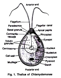
Chlamydomonas is unicellular, motile green algae. The thallus is represented by a single cell. It is about 20 p,-30|i in length and 20 µ in diameter. The shape of thallus can be oval, spherical, oblong, ellipsoidal or pyriform. The pyriform or pear shaped thalli are common, they have narrow anterior end and a broad posterior end (Fig. 1).
The structure of thallus can be divided into following parts:
Cell Wall:
The cell is surrounded by a smooth, thin and firm cell wall made of cellulose. The cell wall at the anterior end is extended to make apical papilla. In some species the outer pectose layer dissolves in water medium to make gelatinous layer outer to cell wall.
The detailed structure of cell wall shows that it is multilayered and is made of cellulose fibrils. Inner to the wall lies the plasma lemma (plasma membrane). It is made of two membranes separated by an opaque zone.
Cytoplasm:
The cytoplasm is present in thallus between the cell wall and the chloroplast. The cytoplasmic structure includes the nucleus, mitochondria, endoplasmic reticulum, dictyosomes, ribosomes etc. The thallus contains single large, dark nucleus lying inside the cavity of the cup shaped chloroplast. The dictyosomes or Golgi bodies are found near the nucleus and they do not possess large vesicles.
Each cell contains two contractile vacuoles located at the base of flagella in a plane at right angle to them. The contractile vacuoles are excretory or osmoregulatory in function. They regulate the water contents of the cell by the process of osmosis. The thallus contains 80S ribosomes while 70S ribosomes characteristic of prokaryotic cells are present in chloroplast (Fig. 2).
Flagella:
The anterior part of thallus bears two flagella. Both the flagella are whiplash or acronematic type, equal in size. The flagella are mostly longer than the thallus but in some species they can be equal or shorter than the thallus. Each flagellum originates from a basal granule or blepharoplast and comes out through a fine canal in cell wall.
Neuro-motor Apparatus:
In some species of Chlamydomonas e.g., C. nasuta, a sensitive neuro-motor apparatus is present. It controls movement of thallus in response to light, chemical and other stimuli.
The neuro-motor apparatus consists of two basal granules or blepharoplasts from which the flagella originate, a transverse cytoplasmic fibre paradesmos which connects two blepheroplasts, a cytoplasmic fibre rhizoplast connecting one blepheroplast with the centrosome and a small delicate fibre connecting centrosome with nucleolus (Fig. 2, 3).
Chloroplast:
In Chlamydomonas generally a large, cup shaped parietal chloroplast is present in cytoplasm (Fig. 4A). But the chloroplasts can be of various shapes in different Chlamydomonas species (Fig. 4B, C).
The chloroplast is ‘H’ shaped in C. bicilliata, reticulate in C. reticulata, parietal in C. mucicola stellate in C. arachne and axile in C. steinii, the chloroplast is generally associated with pyrenoid covered with starch plates, but sometimes pyrenoids can be more than one. The pyrenoids are two in C. debaryana and many in C. gigantae.
The pyrenoids are concerned with synthesis of starch. In chloroplast there are 2-6 thylakods which join to form a granum.
Stigma or Eyespot:
The anterior side of the chloroplast contains a tiny spot of orange or reddish colour called stigma or eyespot. It is photoreceptive organ concerned with the direction of the movement of flagella. The eye spot is made of curved pigmented plate. The plate contains 2-3 parallel rows of droplets or granules containing carotenoids (Fig. 5).
Reproduction in Chlamydomonas:
The reproduction in Chlamydomonas is both asexual and sexual.
Asexual Reproduction:
It takes place by following methods:
(A) By zoospores- The zoospore formation takes place during favourable conditions. The zoospore formation takes place as follows:
The protoplast contracts and gets separated from the cell wall. The parent cell loses flagella or in some species of Chlamydomonas flagella are absorbed. The contractile vacuoles and the neuro-motor apparatus disappear. The protoplasm divides longitudinally by simple mitotic division forming two daughter protoplasts.
The second longitudinal division of protoplasm takes place at right angle to the first, thus making four daughter chloroplasts. Sometimes the protoplasm may further divide to make 8-16-32 daughter protoplasts. The pyrenoids and initials of neuro-motor apparatus also divide. The contractile vacuoles also develop in daughter protoplasts. Each daughter cell develops cell wall, flagella and transforms into zoospore (Fig. 6).
The zoospores are liberated from the parent cell or zoosporangium by gelatinization or rupture of the cell wall. The zoospores are identical to the parent cell in structure but smaller in size. The zoospores simply enlarge to become mature Chlamydomonas. Under favourable conditions the formation of zoospores can take place every 25 hours.
(ii) By Aplanospores:
The aplanospores are formed slightly under unfavorable conditions e.g., in C. caudata. The parent cell loses flagella.
The protoplast rounds off and secretes a thin wall outside but does not develop Fig. 7. (A) Parent cell, (B) Aplanospore formation, (C) Hypnos pore flagella. These non-motile structures are called aplanospores. On approach of favourable conditions aplanospores may germinate either directly or divide to produce zoospores (Fig. 7 A, B).
(iii) By Hypnospores:
In extreme unfavorable conditions the protoplast develops thick wall and the structure developed is called Hypnos pore e.g., in C. nivalis. The hypnospores also germinate like aplanospores on approach of favourable conditions. (Fig. 7 C).
(iv) Palmella Stage:
The palmella stage is formed under unfavorable conditions as shortage of water, excess of salts etc. The protoplast of parent cell divides to make many daughter protoplasts but they do not form zoospores. The parent cell wall gelatinizes to make mucilaginous sheath around daughter protoplasts. The daughter protoplasts also develop gelatinous wall around themselves but do not develop flagella.
These protoplast segments are called palmellospores. The division and red visions of these protoplast ultimately forms amorphous colony with indefinite number of spores and it is called palmella stage (Fig. 8). When favourable conditions return the gelatinous wall is dissolved, palmellospores develop flagella, and the spores ire released to make new thalli.
Sexual Reproduction:
The sexual reproduction in Chlamydomonas can be isogamous, anisogamous or oogamous. he thallus can be homothallic i.e., both types of gametes are produced in same thallus e.g., C. mogama and C. media or can be heterothallic i.e., (+) and (-) gametes come from different parents, he gametes may be naked and called gymnogametes e.g., C. debaryana or covered by cell wall id called calyptogametes e.g., C. media.
(i) Isogamy:
Most of the Chlamydomonas species are isogamous in nature. In isogamous reproduction the fusion of gametes, which are similar in size, shape and structure, take place. These gametes are morphologically similar but physiologically dissimilar.
In many isogamous species the vegetative cells may directly function as gametes without undergoing any division e.g., in C. snowiae (Smith, 1955), this fusion is called as hologamy. The thalli shed their walls and function as gametes.
The two gametes come close to each other by their anterior ends and later fusion proceeds to lateral sides (Fig. 9A-D). The fusion product is quadri flagellate and bi-nucleate structure with two pyrenoids and two eye spots. The quadri flagellate zygote remains motile for several hours to few days. (Fig. 9 E, F).
In C. eugametos, the vegetative cells do not shed their walls, after union the contents of one gamete enter into another gamete as such. According to Chapman (1964) the isogamous reproduction takes place by production of 8, 16 or 32 bi-flagellated gametes. The process takes place as follows (Fig. 10). The vegetative thallus functioning as gametangium comes to rest and loses its flagella.
The protoplast withdraws itself from the cell wall. The protoplast divides by repeated longitudinal mitotic divisions to produce 8-16-32 or 64 daughter protoplasts. Each daughter protoplast develops a pair of flagella and transforms into gamete. The gametes are liberated by breaking the wall of gametangium. The flagella of gametes are covered by agglutins and secrete a hormone called gamone.
These chemical substances are involved in the recognition of gametes of the opposite strains. In heterothallic species (+) and (-) strain gametes cluster together and this phenomenon is called clumping. The gametes of opposite strain fuse by anterior end i.e., apical fusion or laterally i.e., lateral fusion (Fig. 10). The paired gametes move away from the clump.
The wall at the place of contact dissolves and fertilization takes place in two steps—plasmogamy and karyogamy. In plasmogamy the fusion of cytoplasm and in karyogamy the fusion of nuclei takes place. After fertilization a quadriflagellate zygote is formed. The zygote later on loses flagella and gets covered by a thick wall and is now called zygospore.
(ii) Anisogamy:
In anisogamous reproduction the gametes are unequal in size. The male gametes or microgametes are smaller, the female gametes or macrogainetes are larger e.g., in C. braunii and C. suboogama. The macrogametes are formed in female gametangium in which the protoplast divides to make 2 to 4 gametes only (Fig. 11 A, C).
The microgametes are formed in male gametangium where the protoplast divides to make 8-16 gametes (Fig. 11 B, D). The microgametes are more active than macrogametes. The microgametes come close to the macrogamete, the protoplast of microgamete enters into macrogamete and after fusion a diploid zygote is formed. The zygote secretes a thick wall and transforms into zygospore (Fig. 11 E-H).
(iii) Oogamy:
The oogamous sexual reproduction takes place in C. coccifera and C. ooganum. The vegetative thallus functioning as female cell withdraws its flagella and directly functions as non-motile macrogamete or egg. The female gamete contains many pyrenoids (Fig. 12A, B).
The microgametes are formed by four divisions of protoplast as in case of anisogamous reproduction (Fig. 12 C, D). The microgamete reaches the female gamete and unites by anterior ends. The contact wall between the two dissolves. After plasmogamy and karyogamy a diploid zygote is formed (Fig. 12 E-G). The zygote secretes a thick wall and transforms into zygospore.
Zygote/Zygospore:
The zygote is resting diploid spore. The zygote secretes a thick wall which is smooth or ornamented. The zygote accumulates large amount of oils and starch. The zygospores are red in colour due to the presence of haematochrome. the zygospore survives long period of unfavorable conditions and germinates on approach of favourable season.
When the resting period is over and the favourable conditions reappear the zygospore germinates. Its diploid nucleus divides by meiosis to make four haploid nuclei. The four daughter protoplasts, each with one haploid nucleus, form four haploid zoospores or meiozoospores.
Each zoospore contains neuro-motor apparatus, eye spot, two flagella and contractile vacuoles. In 4 zoospores two may be of (+) type and two (-) type in heterothallic forms. The number of meiospores per zygospore are 8 in C. reinhardtii or 16-32 in C. inter-media (Fig,13 A-D, 14, 15).


