Here is a term paper on the ‘Cell’ for class 9, 10, 11 and 12. Find paragraphs, long and short term papers on the ‘Cell’ especially written for school and college students.
Term Paper on the Cell
Term Paper Contents:
- Term Paper on the Introduction to Cell
- Term Paper on the Cell Theory
- Term Paper on the Shape and Size of a Cell
- Term Paper on the Prokaryotic Cell
- Term Paper on the Eukaryotic Cell
- Term Paper on the Structure of an Animal Cell
- Term Paper on the Structure of a Plant Cell
- Term Paper on the Overview of a Cell
1. Term Paper on the Introduction to Cell:
All living organisms on Earth have originated from cells. The cells are the basic building units of all living things similar to the bricks in walls. They provide structure for the body, take in nutrients from food, convert those nutrients into energy, and carry out specialized functions to keep the body functioning properly. Cells also contain the individual’s hereditary material and can make copies of themselves.
All living organisms are made up of cells. Some organisms are made only of one cell and hence, are called as unicellular organisms, e.g. bacteria. Unicellular organisms are capable enough to survive and perform all functions properly which shows a cell’s capability to exist independently.
Due to this, a cell is called the fundamental and structural unit of life. Some organisms are made of many cells. These are called as multicellular organisms. Plants and animals are good examples of the multicellular organisms.
In multicellular organisms, cells unite to form larger structures called tissues. The tissues have cells with similar function and shape which unite to form organs. Numbers of organs are required to carry a function which is called as a system in an organism.
The cells group together to form the tissues of the stomach and eventually the entire digestive system. A cell carries out nutrition, respiration, excretion, transportation and reproduction; the way an individual organism does.
Several cells of one kind that interconnect with each other and perform a shared function form tissues; several tissues combine to form an organ (your stomach, heart, or brain); and several organs make up an organ system (such as the digestive system, circulatory system, or nervous system). Several systems that function together form an organism (like a human being).
2. Term Paper on the Cell Theory:
The first person to see a cell was Robert Hooke. He used a very primitive microscope, but when he was looking at cork cells under the microscope he saw cells for the first time. The shape of the cells reminded him of the monk monasteries and so he dubbed them as “cells.”
In 1665, Robert Hooke discovered the cellular composition of a cork and introduced the word cell to science.
The first person to see living cells was Van Leeuwenhoek, a microscope builder. He named them as “Animalcules”. The Cell Theory was developed from three German scientist’s discoveries. They are Matthias Schleiden, Theodor Schwann, and Rudolph Virchow.
In 1838, a German botanist Matthias Schleiden discovered that all the plants were composed of cells. Then only a year later a German zoologist, Theodor Schwann, discovered that all the animals were composed of cells. Later in 1855, a German physician named Rudolph Virchow was doing experiments with some diseases when he found that all cells come from other existing cells.
Cell theory is as follows:
1. The cell is the fundamental unit of structure and function in living things.
2. All organisms are made up of one or more cells.
3. Cells arise from other existing similar cells through cellular division.
3. Term Paper on the Shape and Size of a Cell:
There is an incredible range of cell shapes, functions and sizes. Prokaryotic cells are much simpler than eukaryotic cells. They have few internal components and a little genetic information. All prokaryotic cells are single cell bacteria and are thought to have been some of the first organisms to exist. The more complex eukaryotic cells make up all other living organisms which have evolved from prokaryotic cells over a period of time.
Prokaryotic cells are the smallest cells, typically a few micro-meters (μm) long. For example, an Escherichia coli (often referred to as E. coli) bacterium is typically 2-3 µm long.
Mammalian red blood cells are among the smallest eukaryotic cells, about 8μm in diameter and have a distinctive bi-concave shape in humans. It is interesting to discover that the cell shape can be very different. For example the red blood cells of a sheep are nearly spherical and those of a camel are elliptical.
An Amoeba is a relatively large single celled organism, typically 10 to 100 µm across.
The largest isolated single cell is the egg of an ostrich.
The cells have variety of shapes depending on the origin and location in an organism. Bacterial cells are of many types. Some are round shaped named as coccus; rod-shaped named as baccilus; chain shaped named as streptococcus and spiral shaped are called spirochete.
Plant cells vary greatly in their shape. They may be disc-like, polygonal, columnar, cuboid, thread like, or even irregular. The shape of the cell may vary with the function they perform. Nerve cells are the longest cells in the animals.
Figure 1.7: Various Cell Shapes in Prokaryotes
4. Term Paper on the Prokaryotic Cell:
There are four main structures shared by all prokaryotic cells, bacterial or Archaean:
1. The plasma membrane
2. Cytoplasm
3. Ribosomes
4. Genetic material (DNA and RNA)
Some prokaryotic cells also have other structures like the cell wall, pili (singular pillus), and flagella (singular flagellum). Each of these structures and cellular components play a critical role in the growth, survival, and reproduction of prokaryotic cells.
1. Prokaryotic Plasma Membrane:
Prokaryotic cells can have multiple plasma membranes. Prokaryotes known as “gram-negative bacteria” for example, often have two plasma membranes with a space between them known as the periplasm. As in all cells, the plasma membrane in prokaryotic cells is responsible for controlling what gets into and out of the cell.
A series of proteins stuck in the membrane (poor fellas) also aid prokaryotic cells in communicating with the surrounding environment. Among other things, this communication can include sending and receiving chemical signals from other bacteria and interacting with the cells of eukaryotic organisms during the process of infection.
Infection is the kind of thing that you don’t want prokaryotes doing to you. Keep in mind that the plasma membrane is universal to all cells, prokaryotic and eukaryotic. Because this cellular component is so important and so common, it is addressed in great detail in its own in Depth subsection.
2. Prokaryotic Cytoplasm:
The cytoplasm in prokaryotic cells is a gel-like, yet fluid, substance in which all of the other cellular components are suspended. It is very similar to the eukaryotic cytoplasm, except that it does not contain organelles. Recently, biologists have discovered that prokaryotic cells have a complex and functional cytoskeleton similar to that seen in eukaryotic cells.
The cytoskeleton helps the prokaryotic cells to divide and help the cell to maintain its plump, round shape. As is the case in eukaryotic cells, the cytoskeleton is the framework along which particles in the cell, including proteins, ribosomes, and small rings of DNA called plasmids, move around. The “highway system” suspended in Jello (for cells).
3. Prokaryotic Ribosomes:
Prokaryotic ribosomes are smaller and have a slightly different shape and composition than those found in eukaryotic cells, bacterial ribosomes, for instance, have about half of the amount of ribosomal RNA (rRNA) and one third fewer ribosomal proteins (53 vs. ∼ 83) than eukaryotic ribosomes have. Despite these differences, the function of the prokaryotic ribosome is virtually identical to the eukaryotic version. Just like in eukaryotic cells, prokaryotic ribosomes build proteins by translating messages sent from DNA.
4. Prokaryotic Genetic Material:
All prokaryotic cells contain large quantities of genetic material in the form of DNA and RNA. Because prokaryotic cells, by definition, do not have a nucleus, the single large circular strand of DNA containing most of the genes needed for cell growth, survival, and reproduction is found in the cytoplasm.
The DNA Tends to Look like a Mess of String in the Middle of the Cell:
Transmission electron micrograph image source Usually, the DNA is spread throughout the entire cell, where it is readily accessible to be transcribed into messenger RNA (mRNA) that is immediately translated by ribosomes into protein. Sometimes, when biologists prepare prokaryotic cells for viewing under a microscope, the DNA will condense in one part of the cell producing a darkened area called a nucleoid.
As in eukaryotic cells, the prokaryotic chromosome is intimately associated with special proteins involved in maintaining the chromosomal structure and regulating gene expression. In addition to a single large piece of chromosomal DNA, many prokaryotic cells also contain small piece of DNA called plasmids.
These circular rings of DNA are replicated independently of the chromosome and can be transferred from one prokaryotic cell to another through pili, which are small projections of the which are small projections of the cell membrane that can form physical channels with the pili of adjacent cells.
Main Cellular Components of Prokaryotes:
The following is the description of the main cellular components of prokaryotes:
1. Cell Wall:
Nearly all prokaryotes have a protective cell wall that prevents them from bursting in an aqueous environment. Cell walls contain a combination of the major organic molecules— proteins, carbohydrates and lipids. The bacteria have a unique molecule called peptidoglycan in their cell wall.
The glycocalyx is a layer present in some bacteria, located outside the cell wall.
There are two types of glycocalyces:
i. Slime layers and
ii. Capsules.
The slime layers help bacteria to stick to things and protect them from drying out, particularly in hypertonic environments. Capsules also allow bacteria to stick to things, and have the added benefit of helping encapsulated bacteria hide from the host’s immune system.
3. Plasma Membrane:
The plasma membrane is a double-layer of phospholipids with associated proteins and other molecules. It is essentially the “bag” that holds all of the intracellular material and regulates the movement of the materials in and out of the cell.
4. Cytoplasm:
Cytoplasm is the fluid that fills a cell, also called as cytosol. The cell organelles are suspended in the cytosol.
5. Mesosomes:
A special membranous structure is called as the mesosome which is formed by the extensions of plasma membrane into the cell. These extensions are in the form of vesicles, tubules and lamellae. They help in the cell wall formation, DNA replication and distribution to daughter cells.
They also help in respiration, secretion processes, to increase the surface area of the plasma membrane and enzymatic content. In some prokaryotes like cyanobacteria, there are other membranous extensions into the cytoplasm called as the chromatophores which contain the pigments.
6. Nucleus:
i. Cell Nucleus:
The cell nucleus acts like brain of the cell. It helps in controlling eating, movement, and reproduction of the cell. The nucleus is not always in the center of the cell. It will be a big dark spot somewhere in the middle of the cytoplasm (cytosol). In some cells, nucleus is missing.
Biology breaks cell types into eukaryotic (those with a defined nucleus) and prokaryotic (those with no defined nucleus). You may have heard of chromatin and DNA. DNA is highly coiled structure embedded in a region in cytoplasm called the nucleoid in prokaryotes.
ii. Plasmids:
In addition to the bacterial chromosome, the bacteria may also contain one or more plasmids. A plasmid is a non-essential piece of the DNA that confers an advantage to the bacteria, such as antibiotic resistance, virulence (the ability to cause disease) and conjugation (a bacterium’s ability to share its plasmids with other bacteria).
7. Cell Extensions:
There are several different types of cell extensions associated with the bacteria, all are made of delicate protein strands.
Bacterial cell extensions include:
i. Flagella:
They are specialized cytoskeletal structures, which are long whip-like or hair-like extensions that help the bacteria to move. Bacterial flagellum is composed of three parts – filament, hook and basal body. The filament is the longest portion and extends from the cell surface to the outside.
ii. Cilia:
They are shorter and more numerous than flagella. In moving cells, the cilia wave in unison and move the cell forward. Paramecium is a well-known ciliated protozoan.
iii. Fimbriae:
They allow bacteria to adhere to the target host cells, hence play a major role in bacterial virulence.
iv. Conjugation Pili:
They are the tubes used to transfer the plasmids from the donor to recipient bacteria.
8. Ribosomes:
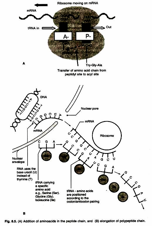
Ribosomes are the protein builders or the protein synthesizers of the cell. Ribosomes are special because they are found in both prokaryotes and eukaryotes. While a structure such as a nucleus is only found in eukaryotes, every cell needs ribosomes to manufacture the proteins.
Since there are no membrane-bound organelles in prokaryotes, the ribosomes float free in the cytosol. Prokaryotic cells have ribosomes made of 50-S and 30-S subunits. They are about 15 nm by 20 nm in size. Several ribosomes may attach to a single mRNA and form a chain called the polyribosomes or polysome. The ribosomes of a polysome translate the mRNA into proteins.
9. Inclusion Bodies:
There are numerous inclusion bodies, or granules, in the bacterial cytoplasm. These bodies are never enclosed by a membrane and serve as storage vessels. Glycogen, which is a polymer of glucose, is stored as a reserve of carbohydrate and energy. Volutin, or metachromatic granules are examples which store the phosphate and energy.
5. Term Paper on the Eukaryotic Cell:
A cell is defined as eukaryotic if it has a membrane-bound nucleus. Any organism composed of eukaryotic cells is also considered a eukaryotic organism. All the organisms we can see with the naked eye are composed of one or more eukaryotic cells, with the most having multiple eukaryotic cells.
Here are some examples of the eukaryotes:
1. Animals
2. Plants
3. Fungi (mushrooms, etc.)
4. Protists (algae, plankton, etc.)
Most plants, animals, and fungi are composed of many cells and are, for that reason, aptly classified as multicellular, while most protists consist of a single cell and are classified as unicellular. All eukaryotic cells have a nucleus, genetic material, a plasma membrane, ribosomes, cytoplasm, including the cytoskeleton. Cells also have other membrane-bound internal structures called the organelles. Organelles include mitochondria, Golgi bodies, lysosomes, endoplasmic reticulum and vescicle.
Organelles of an Eukaryotic Cell:
1. The Nucleus:
Nucleus is like the brain of a body, it controls the cell.
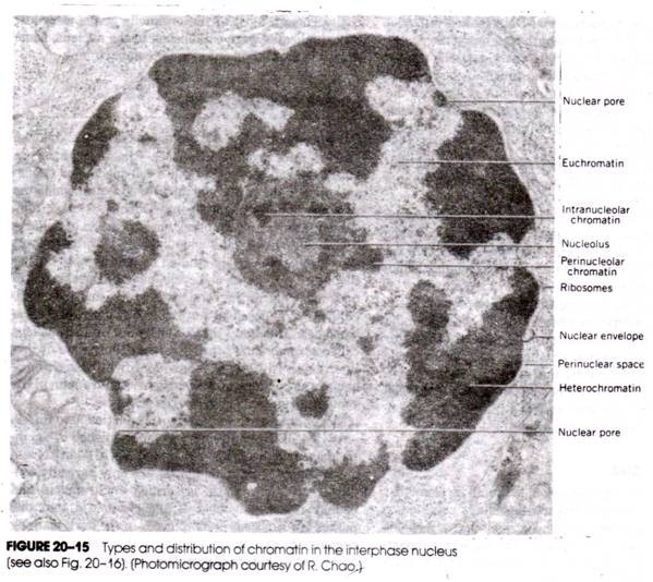
It contains long but thin molecules of DNA (deoxyribonucleic acid) which contains the information needed by the cell to grow, reproduce and function. The nucleus is basically a large membranous sac which protects the cell’s DNA from damage.
It contains a small round body called nucleolus that holds the nucleic acids and proteins. The nuclear membrane has pores through which the contents of the nucleus communicate with the rest of the cell. The nuclear membrane tightly controls and regulates the passage of materials from in and out of nucleus.
Chromosomes:
Chromosomes are structures of proteins and DNA which are sometimes present in the nucleus as condensed chromatin. Chromosomes are single pieces of DNA along with the genes, proteins, and nucleotides.
Chromatin is a condensed package of chromosomes that basically allows all the DNA to fit inside the nucleus. The DNA inside the nucleus is also closely associated with a large protein complex called histone. Along with the nuclear membrane, histones help control which messages are sent from the DNA to the rest of the cell.
2. Plasma Membrane:
Ail prokaryote and eukaryote cells have plasma membranes. The plasma membrane (also known as the cell membrane) is the outermost cell surface, which separates the cell from the external environment.
The plasma membrane is composed primarily of proteins and lipids, especially phospholipids. The cell membrane is not a solid structure. It is made up of millions of smaller molecules that create a flexible and porous container. Proteins and phospholipids are the bricks of the membrane structure.
Fluid Mosaic Model:
Scientists use the fluid mosaic model to describe the organization of phospholipids and proteins. The model shows that the phospholipid molecules are shaped with a head and a tail region. The head section of the molecule likes water (hydrophilic) while the tail does not (hydrophobic).
As the tails want to avoid water, they tend to stick to each other and let the heads face the watery (aqueous) areas inside and outside of the cell. The two surfaces of molecules create the lipid bilayer.
Membrane Protein:
Many proteins are present in the lipid bilayer. Some are permanently attached whereas others are only attached temporarily. There are proteins attached to the inner or outer layer of the membrane whereas the trans-membrane proteins pass through the entire structure.
The trans-membrane proteins that cross the bilayer are very important in the active transport of ions and small molecules. A few ions or molecules are transported across the membrane against their concentration gradient, i.e., from lower to the higher concentration.
Such a transport is an energy dependent process, in which ATP is utilized and is called active transport, e.g., Na+/K+ Pump. Many molecules can move across the membrane without any requirement of energy and this is called as the passive transport. The major reason for this is osmosis.
3. Cell Wall (Structures Present in Plants, Algae and Some Fungi):
Like the cell walls in bacteria, fungi, algae, and some Archaea (single-celled organisms), plant cell walls are found just outside the plasma membrane and provide structure and protection to the cells.
Plant cell walls also have small openings, called the plasmodesmata, which allow the cells to communicate with adjacent cells. In fungi, the cell wall contains a polysaccharide called chitin. The plant cells, in contrast, have no chitin; their cell walls are composed exclusively of the polysaccharide cellulose.
Cell walls provide a support and help cells resist mechanical pressures, but they are not solid, and allow materials to pass through the membrane. Cell walls are not as selectively permeable (semi-permeable), as plasma membranes are.
4. Cytoplasm and Cytoskeleton:
The cytoplasm is a gel-like, yet fluid, substance in which all of the other cellular components are suspended, including all of the organelles. The underlying structure and the function of the cytoplasm, and of the cell itself, is largely determined by the Cytoskeleton (Cyto means “Inside the cell”) is a protein framework along which particles in the cell, including proteins, ribosomes, and organelles, move around.
5. Ribosomes:
A cell require protein and a cell obtains its protein from ribosomes. Ribosomes are found in both prokaryotes and eukaryotes. Ribosomes are the protein factories of the cell made up of proteins and ribosomal RNA. Ribosomes are found in many places around a eukaryotic cell floating in the cytosol. Eukaryotic ribosomes are larger and have a slightly different shape and composition than those that are found in prokaryotic cells.
In eukaryotes, ribosomes are 80S, i.e. made of 60-S (large) and 40-S (small) subunits. Here ‘S’ (Svedberg’s Unit) stands for the sedimentation coefficient; it indirectly is a measure of density and size.
6. Endoplasmic Reticulum:
In eukaryotes there are many membrane bound bodies called as ‘organelles’. Endoplasmic reticulum (which is also referred to as ER) is an organelle. It is a series of membranes extending throughout the cytoplasm of the eukaryotic cells.
It is a network or reticulum of tiny tubular structures scattered in the cytoplasm.
Hence, ER divides the intracellular space into two distinct compartments, i.e.,
i. Luminal (inside ER) and
ii. Extra luminal (cytoplasm) compartments.
The cisternae are spaces within the folds of the ER membranes.
There are two kinds of endoplasmic reticulum, viz.
a. Smooth ER and
b. Rough ER.
The ER is studded with the sub microscopic bodies called ribosomes. This type of ER is referred to as rough ER (RER). It is the site of the protein synthesis and secretion.
Some ER does not contain ribosomes on its surface and hence they are called as smooth ER (SER). In animal cells lipid-like steroidal hormones are synthesised in SER. The ER is the site of protein synthesis in a cell.
7. Golgi Complex:
The Golgi body (or Golgi apparatus) is an organelle which looks like a series of flat sacs, curled at the edges. It was discovered by Camillo Golgi in 1898. In the Golgi body, the cell’s proteins and lipids are processed and packaged before being sent to their final destination. To accomplish this function, the outermost sac of the Golgi body often bulges and breaks away to form drop-like vesicles known as secretory vesicles.
Golgi complex consist of many flat, disc-shaped sacs or cisternae. The golgi cisternae are arranged near the nucleus with a convex forming face called Cis and concave a maturing face called Trans.
8. Lysosomes:
Lysosomes are small sac-like structures arising from Golgi complex. They are small spheres of phospholipids originated from the Golgi bodies. They are responsible for breaking down the cellular debris and material taken into the cell through the process of phagocytosis (the cells swallowing up of things). The interior of a lysosome contains many enzymes and is slightly acidic so that material can be digested without harming the rest of the cell.
9. Vesicles:
Vacuoles are storage bubbles found in the cells. They are found in both the animal and the plant cells but are much larger in plant cells. Vacuoles store food or any variety of nutrients a cell might need to survive. They can even store waste products so the rest of the cell is protected from contamination. Eventually, those waste products would be sent out of the cell.
The structure of vacuoles is fairly simple. There is a membrane that surrounds a mass of fluid. In that fluid are the nutrients or waste products. In plant cells, the vacuoles are much larger than in animal cells.
Plants may also use vacuoles to store water. Those tiny water bags help to support the plant. In Amoeba, the contractile vacuole is important for excretion. In many cells, as in protists, food vacuoles are formed by engulfing the food particles.
10. Mitochondria:
Mitochondria are known as the powerhouses of the cell as they produce cellular energy in the form of ATP. They are organelles that act like a digestive system which takes in nutrients, breaks them down, and creates energy rich molecules for the cell. Mitochondria are small organelles floating free throughout the cell.
Some cells have a several thousand mitochondria while others have none. Muscle cells need a lot of energy so that they have loads of mitochondria. They are made up of two membranes. The outer membrane that covers the organelle and contains it like a skin. The inner membrane that folds over many times and creates layered structures called cristae.
The fluid contained in the mitochondria is called the matrix. Mitochondria are special because they have their own ribosomes and DNA floating in the matrix. There are also structures called the granules which may control concentrations of ions. Cell biologists are still exploring the activity of granules.
11. Plastids:
These are somewhat similar to mitochondria; in appearance. Plastids are found in plant cells. They are of two types, chromoplast and leucoplast. Colourful plastids are called as the chromomplast and colourless plastids are called as the leucoplast. Chloroplast is green in colour and is found in green parts of the plants. Plastids too have their own DNA and ribosome.
i. Leucoplasts:
They are responsible for storing food; such as carbohydrates, protein and lipid. Chromoplasts impart various colours to the plant parts. A leaf of a plant is green in colour because of chloroplast. Chloroplast is the site for photosynthesis which involves water, air and sunlight that is converted to glucose. This is the food for all the plants.
ii. Chloroplast:
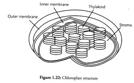
Many plant and algae cells contain small, green organelles called chloroplasts. These organelles are responsible for converting the energy from the sun into chemical energy, usually in the form of glucose, through the elaborate process of photosynthesis.
Chloroplasts are green because they contain large amounts of the green pigment called as chlorophyll bound to proteins embedded in the internal stacks of membranes called the thylakoids.
The sunlight is captured by chlorophyll molecules and transferred throughout the thylakoid membranes to give off energy. This energy is used to break down carbon from carbon dioxide in the air to make sugar. Generally, in any given plant, only some of the cells will contain chloroplasts.
12. Cilia and Flagella:
Flagella and cilia are the extensions of the cell membrane that are lined with the cytoskeleton. Flagella are generally much longer than cilia, like whips, but there are often hundreds or more cilia than flagella on a given cell surface. Flagella are primarily responsible for the cell movement.
Cilia and Flagella are covered with the plasma membrane. Their core is called as the axoneme, possesses a number of microtubules running parallel to the long axis. The axoneme usually has nine pairs of doublets of radially arranged peripheral microtubules, and a pair of centrally located microtubules.
Such an arrangement of axonemal microtubules is referred to as the 9+2 array. The central tubules are connected by bridges and are also enclosed by a central sheath, which is connected to one of the tubules of each peripheral doublet by a radial spoke.
Thus, there are nine radial spokes. The peripheral doublets are also interconnected by the linkers. Both the cilium and flagellum emerge from a centriole-like structure called the basal body.
13. Centrosome and Centriole:
Centrosome is an organelle usually containing two cylindrical structures called centrioles. They are surrounded by amorphous pericentriolar materials. Both the centrioles in a centrosome lie perpendicular to each other. This structure has an organization like the cartwheel.
Centrioles are present to help the cell during the time of the division. A centriole is a small set of microtubules arranged in a specific way. There are nine groups of microtubules. When two centrioles are found next to each other, they are usually at right angles.
The centrioles are found in pairs and move towards the poles (opposite ends) of the nucleus when it is time for the cell division. During the division, you may also see groups of threads attached to the centrioles. Those threads are called the mitotic spindle.
Many membrane bound minute vesicles called microbodies that contain various enzymes, are present in both the plant and animal cells.
6. Term Paper on the Structure of an Animal Cell:
Let us now look at the individual cell organelles to understand their structure and functions:
7. Term Paper on the Structure of a Plant Cell:
8. Term Paper on the Overview of a Cell:
Scientists distinguished all the organisms on the basis of cell structure, organelles and cell characteristics as prokaryotes or eukaryotes. Prokaryotes are single- celled organisms, including bacteria and their bacteria-like cousins called Archaea.
Prokaryotic cells are much simpler than the more evolutionarily advanced eukaryotic cell. Whereas eukaryotic cells have many different functional compartments, divided by membranes, prokaryotes have only one membrane called the plasma membrane.
This membrane encloses all of the cell’s internal contents and is a barrier to the surroundings. If a eukaryotic cell is analogous to a big house with many different rooms, a prokaryotic cell is like a one-room, studio apartment.
The cells of all prokaryotes and eukaryotes possess two basic features:
i. A plasma membrane and
ii. The cytoplasm.
Prokaryotic cells lack a nucleus and have the DNA embedded directly in the cytoplasm as nucleolus, while eukaryotic cells have a nucleus which has nucleoplasm and DNA.
Prokaryotic cells lack internal cellular bodies with membrane (organelles), while eukaryotic cells possess them. Examples of prokaryotes are bacteria and cyanobacteria (formerly known as blue green algae). Examples of eukaryotes are protozoa, fungi, plants, and animals.
Let us see the structure of the cell. Cells are highly organized units composed of living material. Many kinds of prokaryotes and eukaryotes contain a structure outside the cell membrane called cell wall, which protects the cell from the surroundings.
With only a few exceptions, all bacteria have thick, rigid cell walls that give them their shape and maintains size. Among the eukaryotes, fungi and plants have cell walls. Cell walls provide a support and help cells resist mechanical pressures, but they are not solid, so materials are able to pass through rather easily.
The plasma membrane (also known as the cell membrane) is the outermost cell surface, which separates the cell from the external environment. Inside each cell there is a dense membrane bound structure called the nucleus.
The nucleus contains proteins and genetic material deoxyribonucleic acid, or DNA. The DNA is organized into linear units called chromosomes; segments of the chromosomes are referred to as genes. Approximately 100,000 genes are located in the nucleus of all human cells.
Cells also have cytoplasm (or cytosol), a semiliquid gel-like material enclosed by the plasma membrane. Within the cytoplasm of eukaryote cells are a number of membrane-bound bodies called organelles (“little organs”) that provide a specialized function within the cell.
Some of the organelles are Endoplasmic Reticulum (ER), the Golgi complex, Lysosomes, Mitochondria, Micro-bodies and Vacuoles. The prokaryotic cells lack such membrane bound organelles.
Ribosomes are non-membrane bound organelles found in all the cells — both eukaryotic as well as prokaryotic cells. Within the cell, ribosomes are found not only in the cytoplasm but also within the two organelles – chloroplasts (in plants) and mitochondria and on rough ER.
The animal cells contain another non-membrane bound organelle called the centriole which helps in cell division.


