In dicotyledon the vascular bundles are usually more or less in a ring and show different arrangements at the nodes and internodes. The vascular cylinders are generally continuous at the internode and their continuity is interrupted at the nodal region due to the emergence of bundles that terminate either at the leaf bases, axillary buds or stipules etc.
At the node three types of bundles are recognized:
(i) Leaf trace bundle:
The single vascular bundle that connects the leaf base with the main vascular cylinder of stem is designated as leaf trace bundle. In a leaf there may be several leaf trace bundles that collectively are termed as leaf traces.
(ii) Cauline bundle:
The vascular bundles that entirely form the vascular system of stems are known as cauline bundles. Sometimes these bundles anastomose with each other and extend from stem to leaf as leaf traces.
(iii) Common bundle:
The vascular bundles, which run unbranched through a few successive nodes and internodes and ultimately terminate as leaf traces are called common bundles.
The arrangement of vascular tissues at the nodes is more complex than the internodes due to emergence of vascular traces to the leaves, buds, stipules etc., present at the node.
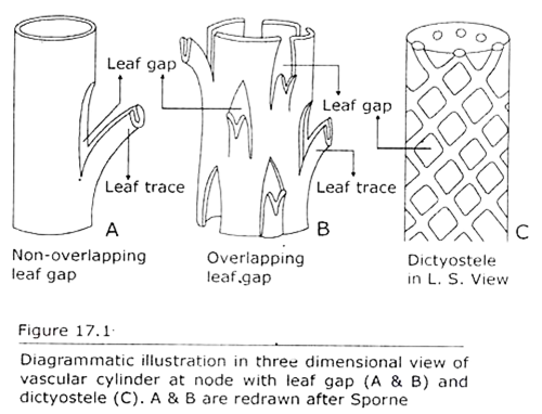
i. Leaf trace and leaf gap (Fig. 17.1A, B & C):
A leaf trace is defined as the cauline part of vascular tissue that departs from the stele of stem towards leaf base. Leaf trace may be a portion of cauline bundle as it occurs within the caulis, that is, stem. Cauline bundles entirely form the vascular system of stems.
Sometimes cauline bundle departs from the stele towards leaf base thus forming leaf trace. Leaf trace is seen in the nodal region of stem. Leaf trace is an independent bundle that may occur through one or more nodes and internodes before bending away from the stem toward the leaf.
A leaf trace originates from the apical meristem of leaf primordium. After complete differentiation it joins with the vascular tissue of stem. Apart from leaf trace other vascular strands entirely occur in the stem. They constitute the cauline (= stem) bundle. They originate from apical meristem of shoot apex. Some cauline bundles may bend towards leaf thus forming leaf trace and these bundles are referred to as common bundles.
In the nodal region, a leaf trace bends away from the vascular cylinder of stem toward the petiole of leaf. From the base of petiole leaf trace extends into leaf blade where the trace forms vascular bundle of leaf. In the stem phloem occurs on the peripheral side of vascular cylinder.
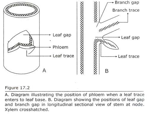
As the leaf trace is bent away from the vascular cylinder toward petiole phloem in the vascular bundle of leaf occurs on the abaxial side of leaf (Fig. 17.2A). As a result the vascular bundle of leaf has an inverted orientation of xylem and phloem in relation to vascular bundle of stem.
A cross-section through node reveals that, there is a break in the vascular cylinder of stem, that is, the vascular cylinder is not in the form of a continuous ring. In this region the vascular tissue is interrupted and parenchyma cells fill the gap. Pith and cortex are continuous through this gap (Fig. 17.6).
Such region is referred to as leaf gap and occurs opposite to leaf trace. A leaf gap is defined as the wide interfascicular region that is located opposite the upper part of leaf trace and is filled up with parenchyma cells through which pith and cortex become continuous.
There exist many variations in the number of leaf gaps and the number of leaf traces in different plants. The variation may be different in the same plant at different levels, e.g. Hypecoum procumbens—the lower node of which has three traces per leaf whereas the upper nodes have single trace per leaf. The variations are subjects of comparative studies and therefore have taxonomic and phylogenetic significance.
Atactostele is the characteristic of monocot stem and the arrangement of vascular strands at the node is highly complex. Comparatively the anatomy at the node of dicotyledonous stem is less complex. To describe the nodal anatomy of stem different terms are used.
In the terminology the term lacuna replaces the term gap. Dicotyledonous nodes are very suitable to study the nodal anatomy and the different terminology used to describe the anatomy of nodes is chiefly based on dicotyledons.
The following types of anatomy at nodes in dicots are recognized:
(a) Unilacunar single-trace node:
This type of node exhibits one leaf trace to a leaf and the leaf trace is associated with one lacuna. Ex. Spiraea. Unilacunar nodes are exstipulate (Figs. 17.3 & 17.4A).
(b) Unilacunar two-trace node:
This type of node has two leaf traces to a single leaf and one lacuna. The two leaf traces are associated with the single lacuna. Ex. Clerodendron (Fig. 17.3).
(c) Trilacunar node:
This type of node has three leaf traces to a leaf and three lacunae. Each trace is associated with single lacuna (Fig. 17.4B). Ex. Salix. Among the three traces, the trace that occurs in the median position with reference to leaf is referred to as median trace. The others are called lateral traces. This type of node has stipules.
(d) Multilacunar node:
This type of node has more than three traces to a leaf and more than three lacunas. Each trace is confronted with single lacuna. Ex. Rumex. Multilacunar node is exhibited in the plants that have sheathing leaf bases.
It is to note that the terms unilacunar-, trilacunar- and multilacunar node are applied with reference to a single leaf. In this sense in unilacunar node each leaf is associated with one lacuna. A node may consist more than one leaf. A cross-section of such node reveals the presence of more than one lacuna and more than one leaf trace.
Such node is to be referred in relation to the number of lacuna(s) associated to a single leaf, that is, in trilacunar node one leaf is associated with three lacunas and in multilacunar node one leaf is associated with several lacunas. In other words, whatever may be the number of leaves present, a node is characterized with reference to the number of lacuna(s) present in a single leaf.
In unilacunar node a leaf has one trace, in trilacunar node three traces diverse to a single leaf and in multilacunar node several traces are present in a single leaf. Usually one gap is associated with one trace but in two-trace unilacunar condition two leaf traces confront to a single gap and in three-trace unilacunar condition three leaf traces are associated to a single gap (Figs. 17.3 & 17.4C).
It is mentioned previously that a leaf gap is identifiable at the node. In internode also leaf gaps may be identified when a leaf trace has oblique course through a part of the internode. Studies on the cross-section of internode, if followed level by level, will reveal position(s) of leaf gap(s) and the continuity between ground tissue and pith.
A definite nodal type generally characterizes a taxon. Unilacunar node is found in Centrospermae and in certain members of Myrtaceae (Eucalyptus), Lauraceae (Laurus) etc. Trilacunar node occurs in Compositae (Chrysanthemum), Salicaceae (Salix), Brassicaceae (Brassica) etc. Multilacunar node is observed in Polygonaceae (Rumex) etc.
The anatomy of the node is regarded an important aspect in the study of phylogeny in dicotyledons, since a definite nodal type characterizes a taxon. According to Sinnott (1914) trilacunar node is primitive among dicotyledons. During evolution it gave rise to unilacunar type by reduction in the number of gaps and traces.
Reduction occurred either by the disappearance of the two lateral gaps with the associated two lateral traces or the two lateral traces became confluent with the median trace. In the latter case a single bundle is formed consisting of three traces. This bundle is associated with a single gap to form unilacunar node.
Trilacunar condition also gave rise to multilacunar type by the formation of more new gaps and traces. Though unilacunar condition is considered as advanced, later studies on nodal anatomy by Bailey (1956), Fahn and Bailey (1957) and others reveal that unilacunar condition is primitive as this type is found in some primitive groups like pteridophyta, fossil gymnosperms like Bennettitales and Cordaitales, Ginkgo and Ephedra.
Unilacunar two-trace node also seems to be primitive as it is represented by extinct Cordaitales and Bennettitales, and extant ferns, conifers, Ephedra and the primitive dicotyledonous genus Austrobaileya etc.
Trilacunar node is widespread and is exhibited in the families like Winteraceae, Meliaceae, Rosaceae and Asteraceae etc. Degeneriaceae and Chenopodiaceae etc. exhibit multilacunar condition. Lepidium latifolium has unilacunar node with several leaf traces. It is regarded as more primitive than the unilacunar one-trace node.
Previously the nodal structures were described on the basis of number of leaf traces that are associated with each leaf, e.g. one-trace, two-trace, three-trace and multi-trace.
Later the nodal anatomy is interpreted on the basis of leaf gaps that are associated with each leaf, e.g. unilacunar, trilacunar and multilacunar. Still later leaf gap and leaf trace —these two aspects were combined to describe a node, e.g. unilacunar-one trace, unilacunar-two trace etc.
Before the discovery of unilacunar two-trace node, trilacunar node was regarded as central type from which unilacunar and multilacunar node arose. It is interpreted that unilacunar node is more advanced than trilacunar node.
Later in the light of above facts it is now interpreted that the evolution of nodal structure proceeded in the following two sequences:
(i) Two trace unilacunar gave rise to trilacunar, which terminated in multilacunar condition;
(ii) Two-trace unilacunar, by the loss of one trace, gave rise to one trace unilacunar that formed trilacunar node by the addition of two new gaps associated with two traces.
The multilacunar condition is derived from the trilacunar type by the addition of more new traces and gaps (Fig.17.5). This evolutionary sequence may be observed in a single family namely Chenopodiaceae. The trilacunar type may also give rise to one trace unilacunar condition.
ii. Branch trace and branch gap:
Branch trace can be defined as the vascular trace that originates from the vascular cylinder of stem and enters to a branch. The position of the origin of a branch is from the axil of a leaf (Fig. 17.4A). In this position the vascular cylinder of stem is discontinuous. Parenchyma occupies this region as observed in a cross-section of stem at node.
This interrupted region of vascular cylinder due to the presence of branch trace is designated as branch gap. Two branch traces are associated to a single leaf gap. Branch trace directly diverges to a branch from the main vascular cylinder of stem without running obliquely through the internode.
The branch traces depart from the main vascular cylinder of stem toward right and left of the median leaf trace. After a short distance the traces coalesce and form a complete stele similar to main vascular cylinder. So the vascular bundle of branch has the same orientation of xylem and phloem in relation to the vascular bundle of stem.
It is previously mentioned that branch develops from axillary bud that originates on the stem at the axil of leaves. From the time of initiation the buds are connected by vascular traces to the vascular strands on the main axis. These vascular traces are referred to as bud trace or branch trace or ramular trace.
Axillary buds form axillary shoots whose first foliar structures are the prophylls. In dicotyledons, usually two branch traces emerge out from the vascular cylinder of stem. In exceptional cases the branch trace may be one (e.g. Peperomia, Cayratia etc.) or more than two. Sometimes medullary bundles may enter into the bud (Ex. Dahlia).
In case of single branch supply the vascular cylinder appears as crescent or horseshoe shaped in cross sectional view with the opening downward. After a short distance the opening closes and the cylindrical stele of the branch is formed. When the branch traces are more than one they coalesce after a short distance, forming a complete stele similar to that of the main axis.
In dicotyledons there occurs two prophylls, which are oriented in such a way that their plane of bisection is parallel with the plane of the axillant leaf. Initially two branch traces supply the prophylls. The branch traces are composed of one or more bundles that later increase in size due to the formation of vascular traces to the other leaves of the branch, situated above. Thus the branch traces, are actually the leaf traces of the axillary shoot.
The continuity of the vascular cylinder of stem is interrupted at the nodal region due to the emergence of branch traces. At this region and above the point of departure parenchyma differentiates instead of vascular tissues. The parenchymatous area in the vascular cylinder of stem at the node immediately above the branch trace is the branch gap through which pith and cortex become continuous.
Branch gap occurs in those vascular plants, which have pith. In pteridophytes, where the vascular cylinder is protostele and devoid of pith, branch gap is absent. So the branch gap in association with leaf gap results in the formation dissected siphonostele.
There exists a definite correlation between leaf—and branch trace. As seen in cross-section of a stem at node a leaf trace occurs on the peripheral side. It is followed by branch trace towards the inner side (Fig. 17.3). A node that bears a single leaf exhibits the following sequences from periphery towards centre —leaf trace, branch trace and leaf gap on the side where the leaf is inserted.
When there are two leaves in each node the above mentioned sequences of leaf—and branch trace and leaf gap are observed on the opposite sides in a cross-section of stem at node. In longitudinal section the positions of branch gap and leaf gap (Fig. 17.2B), and their relation can be observed individually.


