Notes on Meiosis: Features, Division and Significance!
Notes # Features of Meiosis:
Important features of meiosis are presented below:
Two spindle using divisions which reduce the chromosome number from diploid to haploid constitute meiosis. The main function of meiosis is to produce gametes in an organism.
1. Meiosis results in formation of four daughter cells from a single mother cell in each cycle of cell division. In other words, the nuclei divide twice in each cell cycle.
2. Daughter cells are identical to mother cell in shape and size but different in chromosome composition. The daughter cells have haploid chromosome number. The chromosome types also differ in daughter cells due to segregation and recombination.
3. Meiosis occurs in reproductive organs like anthers and ovaries and leads to the production of gametes or spores.
4. The complete process of meiosis consists of two types of division. The first division results in reduction of chromosome number to half and is called reductional division. The second division is like mitotic division.
5. Meiosis results in segregation of chromosomes and genes and their independent assortment. Crossing over and recombination also occur during meiosis.
Notes # Division of Meiosis:
The process of meiosis as indicated earlier, consists of two types of division, viz., first meiotic and second meiotic division. Before initiation of meiosis, there is an interphase which consists of G1, S and G2 phases like mitosis. But here the G2 phase is of very short duration.
The S phase occurs only once in the entire process of meiosis. There is no S phase after first division of meiosis. During S phase 99.7% of the total DNA present in the nucleus is synthesized and remaining 0.3% DNA synthesis takes place during zygotene stage.
A. First Meiotic Division:
The first meiotic division results in reduction of chromosome number in each new cell to just half of the mother cell, therefore, it is referred to as reductional division.
The first meiotic division consists of four different phases, viz:
(1) Prophase,
(2) Metaphase,
(3) Anaphase and
(4) Telophase.
1. First Prophase:
This phase starts after interphase and is of maximum duration. This consists of five sub stages, viz., leptotene, zygotene, pachytene, diplotene and diakinesis.
Important features of these sub stages are briefly discussed below:
a. Leptotene:
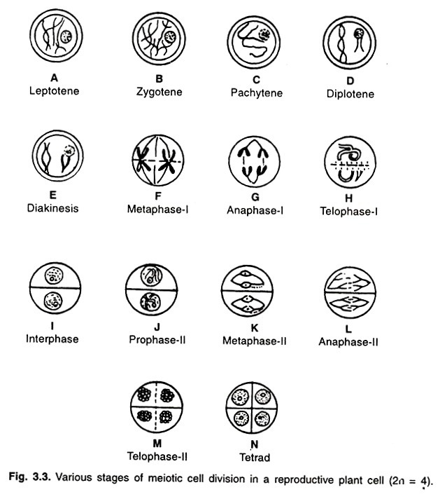
i. Chromosomes look like thin thread under light microscope. They are inter-woven like a loose ball of wool (Fig. 3.3A).
ii. Chromosomes are scattered throughout the nucleus in a random manner.
iii. In some cases, chromomeres are visible on the chromosomes in the form of condensed regions.
iv. RNA and protein syntheses also take place.
b. Zygotene:
i. Homologous chromosomes being to pair.
ii. Chromosomes become shorter and thicker (Fig. 3.3B).
iii. The synthesis of remaining 0.3%. DNA which has not taken place during S phase also occurs during this stage.
iv. Synaptonemal complex also develops during this stage.
c. Pachytene:
i. Chromosomes look like bivalents. Each bivalent has two chromatids. Thus each pair has four chromatids generally known as tetrads.
d. Diakinesis:
i. This stage begins after complete terminilization of chiasmata (Fig. 3.3E).
ii. Chromosomes are further condensed.
iii. Bivalents are distributed throughout the cell.
iv. Nucleolus and nuclear membrane disappear towards the end of diakinesis.
2. First Metaphase:
i. The spindle apparatus gradually organises.
ii. Bivalents are arranged on the equatorial plate (Fig. 3.3F).
iii. The centromere of each chromosome divides longitudinally.
3. First Anaphase:
i. From each bivalent, one chromosome moves towards one pole and the other towards other pole. In other words, one homologous chromosome moves towards one pole and another to opposite pole (Fig. 3.3G).
ii. Sister chromatids of each chromosome remain attached at the centromere.
iii. Homologous chromosomes reach the opposite pole at the end of this phase.
4. First Telophase:
i. Chromosomes uncoil and relax and regrouping of chromosomes occurs (Fig 3.3H).
ii. Nucleolus and nuclear membrane reappear.
iii. Two haploid daughter nuclei are formed.
B. Second Meiotic Division:
The first nuclear division (Meiosis I) results in reduction of chromosome number from diploid to haploid. The second nuclear division (Meiosis II) is required to reduce the number of chromatids per chromosome.
Meiosis II differs from Mitosis in the following three main aspects:
1. The interphase prior to meiosis II is very short. It does not have S period because each chromosome already contains two chromatids.
2. The two chromatids in each chromosome are not sisters throughout. In other words, some chromatids have alternate segments of non-sister chromatids due to recombination.
3. The meiosis II (Meiotic mitosis) deals with haploid chromosome number, whereas normal mitosis deals with diploid chromosome number.
Rest of the features of meiosis II is similar to mitosis. It also consists of prophase, metaphase, anaphase and telophase.
Cytokinesis:
The division of cytoplasm takes place either by cell plate method (in plants) or by furrow method (in animals). The cytokinesis may take place after meiosis I and meiosis II separately or sometimes it may take place at the end of meiosis II only. In maize, it occurs after meiosis I and meiosis II. However, in Trillium cytokinesis occurs only at the end of Meiosis II.
2. The chromosome number looks like haploid number.
3. Nucleolus is present and attached to a chromosome.
4. Formation of chiasma and crossing over take place during pachytene stage (Fig. 3.3C).
Diplotene:
1. Separation of homologous chromosomes begins. It starts at centromere and moves towards the end (Fig. 3.3D).
2. The separating chromosomes are attached at some points. These points are called chiasmata. These chiasmata are terminalized towards the end of diplotene.
3. Chromosomes are further condensed and become still shorter and thicker.
4. Nucleolus decreases in size.
Synaptonemal Complex:
It is a protein framework which is found between paired chromosomes. It consists of one central and two lateral elements. There are transverse filaments on both the sides of central element. The lateral elements are attached to homologous chromosomes (Fig. 3.4).
Synaptonemal complex is considered to be associated with pairing of homologous chromosomes and recombination. However, its origin and exact role in synapsis is still not properly known which requires further research.
Notes # Genetic Control of Meiosis:
It is believed that meiosis is genetically controlled. Various features of meiosis are controlled by genes.
Some of the features of meiosis which are genetically controlled are described below:
1. Synapsis and Exchange:
Synapsis or pairing between homologous chromosomes may depend on the presence of a specific allele. For example, in maize when this allele is absent, synapsis is prevented between all homologous loci. In the absence of synapsis, no exchange occurs between homologous chromosomes and distribution of chromosomes is also irregular during anaphase I.
In Drosophila male, crossing over does not occur because the homologous chromosomes pair only in the heterochromatic region near centromere. Heterochromatin is considered devoid of active genes, hence exchange is prevented. However, the pairing is normal in females.
2. Centromere Behaviour:
The specific behaviour of centromere during meiosis is genetically controlled. At metaphase II, centromeres of sister chromatids lie very close together. But when one of them faces one pole of spindle, the other one automatically faces the opposite pole.
In Drosophila, in the presence of a particular allele, sister centromeres separate early at metaphase II and orient to the spindle independently. As a result, both sister centromeres sometimes orient to the same pole and hence are not distributed to daughter nuclei properly. However, this allele has no effect on mitosis or meiosis I.
3. Spindle Shape:
The shape of spindle is governed by a specific allele. The presence of abnormal alleles changes the shape of spindle in maize and Drosophila during meiosis but not mitosis. A normal allele causes the meiotic spindle to have convergent ends.
With such spindle, all the chromosomes come together in group to the pole and are included in a telophase nucleus. In the presence of abnormal allele, divergent spindles are formed. Such spindles lead to the spread of chromosomes at anaphase in such a way that some are left but of telophase nuclei.
4. Spindle Orientation:
The orientation is usually similar in meiosis I and meiosis II. This leads to formation of four nuclei or cells. When this direction is opposite to that of meiosis I, the result is a cluster of four nuclei or cells.
Notes # Significance of Meiosis:
Meiosis plays a very important role in the biological populations in various ways as given below:
1. It helps in maintaining the chromosome number constant in a species. Meiosis results in production of gametes with haploid (half) chromosome number. Union of male and female gametes leads to formation of zygote which receives half chromosome number from male gamete and half from the female gamete and thus the original somatic chromosome number is restored.
2. Meiosis facilitates segregation and independent assortment of chromosomes and genes.
3. The recombination of genes also takes place during meiosis. Recombination of genes results in generation of variability in a biological population which is important from evolution points of view.
4. In sexually reproducing species, meiosis is essential for the continuity of generation. Because meiosis results in the formation of male and female gametes and union of such gametes leads to the development of zygote and thereby new individual.

