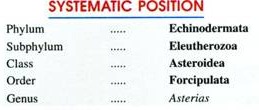
In this article we will discuss about Asterias (Starfish):- 1. History of Asterias 2. Habit and Habitat of Asterias 3. External Features 4. Body Wall 5. Endoskeleton 6. Coelom 7. Digestive System 8. Water Vascular System 9. Locomotion 10. Circulatory System 11. Respiratory and Excretory System 12. Nervous System 13. Sense Organs 14. Reproductive System 15. Life History and Development 16. Regeneration and Autotomy.
Contents:
- History of Asterias
- Habit and Habitat of Asterias
- External Features of Asterias
- Body Wall of Asterias
- Endoskeleton of Asterias
- Coelom in Asterias
- Digestive System of Asterias
- Water Vascular System of Asterias
- Locomotion of Asterias
- Circulatory System of Asterias
- Respiratory and Excretory System of Asterias
- Nervous System of Asterias
- Sense Organs of Asterias
- Reproductive System of Asterias
- Life History and Development of Asterias
- Regeneration and Autotomy of Asterias
Contents
- 1. History of Asterias:
- 2. Habit and Habitat of Asterias:
- 3. External Features of Asterias:
- 4. Body Wall of Asterias:
- 5. Endoskeleton of Asterias:
- 6. Coelom in Asterias:
- 7. Digestive System of Asterias:
- 8. Water Vascular System of Asterias:
- 9. Locomotion of Asterias:
- 10. Circulatory System of Asterias:
- 11. Respiratory and Excretory System of Asterias:
- 12. Nervous System of Asterias:
- 13. Sense Organs of Asterias:
- 14. Reproductive System of Asterias:
- 15. Life History and Development of Asterias:
- 16. Regeneration and Autotomy of Asterias:
1. History of Asterias:
Asterias is commonly known by the name of starfish. The name starfish is somewhat misleading suggesting an organism to be like a star and fish but as Asterias lacks in both the characteristics, therefore, recently it is renamed as sea star. There occur about 150 species of Asterias all of which have different geographical distribution.
Asterias rubens occurs on the English and North European coasts, A. vulgaris is found on the North Atlantic coast of North America, A. forbesi occurs on the eastern sea shore from the Maine to the Gulf of Mexico, A. amurensis is found in the Behring sea, Japan and Korea, and A. tenera occurs on the sea shore from Nova Scotia to New Jersey.
Some other common sea stars are Pentaceros, Astropecten, Asterina, Heliaster, Solaster, Luidia, etc. The following account will give a general idea about the anatomical organisation of the genus Asterias.
2. Habit and Habitat of Asterias:
Asterias is exclusively marine, bottom dwelling or benthonic animal, inhabiting various types of bottom, mainly in the littoral zone where they crawl about or may remain quiescent at time’s, either in the open or more or less concealed. Asterias forbesi is found equally abundant on hard, rocky, sandy or soft bottom, while other species have been found to prefer rocky sea bottoms.
The most species of Asterias are generally solitary but under certain ecological conditions, such as to avoid direct sunlight or excessive drying, many individuals may gather at some place for the purpose of protection. Most of them are nocturnal, remain quiet in day time and become active during night. They move by crawling on the bottom, mostly at a rather slow rate.
All sea stars are carnivorous and feed voraciously on almost any available slow moving or sessile animals, chiefly on polychaetes, crustaceans, molluscs and other echinoderms and even corpses.
Many species of Asterias exhibit various types of biological relationships such as parasitism and commensalism, etc., with the members of different zoological groups. Sea stars, in general, exhibit remarkable power of autotomy and regeneration.
3. External Features of Asterias:
(i) Shape, Size and Colour:
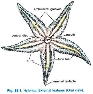
Asterias has a radially symmetrical and pentamerous body. The body consists of a central, pentagonal central disc from which radiate out five elongated, tapering, symmetrical spaced projections, the rays or arms. In some genera, the number of arms may be more than five, for example, there are 7-14 arms in Solaster and more than 40 arms in Heliaster.
The size varies from 10-20 cm in diameter though some forms may be much smaller or longer. The colour is variable having shades of yellow, orange, brown and purple. The body has two surfaces, the upper convex and much darker side is called the aboral or abactinal surface.
The lower surface is flat, less pigmented and is called the oral or actinal surface. The oral and aboral surfaces are not the ventral and dorsal surfaces but correspond to the left and right sides of the bilaterally symmetrical larva. The axes occupied by the arms are known as radii and the regions of the central disc between the arms are inter-radii. A well defined head is entirely absent.
(ii) Oral Surface:
The side of body, which in natural condition remains towards the substratum and contains the mouth or oral opening, is flat and of dark orange to purplish colour, is called oral or actinal surface.
The oral surface bears the following structures:
1. Mouth:
On the oral surface, in the centre of the pentagonal central disc is an aperture, the actinosome or mouth. It is a pentagonal aperture with five angles, each directed towards an arm. The mouth is surrounded by a soft and delicate membrane, the peristomial membrane or peristome and is guarded by five groups of oral spines or mouth papillae.
2. Ambulacral Grooves:
From each angle of the mouth radiates a narrow groove called the ambulacral groove which runs all along the middle of oral surface of each arm.
3. Tube Feet or Podia:
Each ambulacral groove contains four rows of locomotory, food capturing, respiratory and sensory organs called tube feet or podia. The tube feet are soft, thin- walled, tubular, retractile structures provided with terminal discs or suckers. The suckers function as suction cups to afford a firm attachment on the surface to which they are applied.
4. Ambulacral Spines:
Each ambulacral groove is bordered and guarded laterally by 2 or 3 rows of movable calcareous ambulacral spines which are capable of closing over the groove. Near the mouth, these spines often become larger, stouter, assemble in five groups, one at each inter-radius of disc and are called mouth papilla.
Outside the ambulacral spines are three rows of stout immovable spines, beyond which occurs another series of marginal spines along the borders of the arms demarcating the oral from the aboral surface.
5. Sense Organs:
Sense organs include five unpaired terminal tentacles and five unpaired eye spots. The tip of each arm bears a small median, non-retractile and hollow projection, the terminal tentacle. It acts as a tactile and olfactory organ. At the base of each tentacle occurs a bright red photo-sensitive eye spot made up of several ocelli.
(iii) Aboral Surface:
The side of the body, which remains directed upward or towards the upper surface, is convex and of light orange to purplish colour, is called aboral or abactinal surface.
The aboral surface bears following structures:
1. Anus:
A minute circular aperture, called the anus, is situated close to the centre of the central disc of aboral surface.
2. Madreporite:
At the aboral surface of the central disc occurs a flat, sub-circular, asymmetrical and grooved plate called madreporite plate or madreporite between the bases of two of the five arms. The surface of madreporite is marked by a number of radiating, narrow, straight or slightly wavy grooves with pores in them. The madreporite is, thus, a sieve-like porous plate and it leads to the stone canal of water vascular system.
The number of madreporite to an individual though remains one, but the presence of more than one madreporite in some species is due to the increase in number of arms beyond the normal number of five.
The two arms having madreporite between their bases are collectively referred to as a bivium and the other three arms as a trivium. The symmetrical position of madreporite, thus, converts the radial symmetry of Asterias into bilateral symmetry.
3. Spines:
The entire aboral surface is covered with numerous short, stout, blunt, calcareous spines or tubercles. The spines are variable in size and are arranged in irregular rows running parallel to the long axes of the arms. The spines are supported by the irregularly-shaped calcareous plates or ossicles which remain buried in the integument and form the endoskeleton.
4. Papulae or Gills:
Between the ossicles of integument are present a large number of minute dermal pores. Through each dermal pore projects out a very small, delicate, tubular or conical, finger-like or thread-like, thin-walled, membranous and retractile projection called the dermal branchia or gill or papula.
The papulae are hollow evaginations of the body wall and their lumen remains in continuation with the coelom. They are internally lined by coelom. They have respiratory, as well as excretory functions.
5. Pedicellariae:
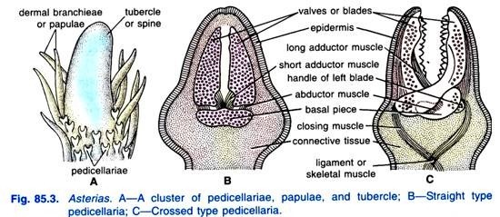
Besides the spines and gills, entire aboral surface is covered by many whitish modified spine-like tiny pincers or jaws called pedicellariae. The oral surface also bears pedicellariae. Each pedicellaria consists of a long or short, stout, flexible stalk having no internal calcareous support.
The stalk bears three calcareous ossicles or plates a basilar piece or plate at the extremity of the stalk and jaws or valves which remain movably articulated with the basilar piece and serrated along their apposed edges. Pedicellariae having three calcareous pieces and a stalk are called forcipulate pedunculate pedicellariae.
Asterias possesses two types of forcipulate pedunculate pedicellariae. viz.:
(i) Straight type and
(ii) Crossed type.
(i) Straight type Pedunculate Pedicellariae:
This type of pedicellariae are simple. Their two jaws are more or less straight and attached basally to the basal piece. When closed they remain parallel and meet throughout their length. The two jaws work against one another like the blades of a force with the help of three pairs of muscles. There are two pairs of adductor muscles for closing the jaws and a pair of abductor muscles for opening them.
(ii) Crossed type Pedunculate Pedicellariae:
In the crossed type of pedicellariae, the basal ends of the two jaws cross each other like the mandibles of a crossbill, so that the basal piece is enclosed between their crossed portions. In this type of pedicellariae, the jaws are also operated by two pairs of adductor muscles and one pair of abductor muscles.
Certain other pedicellariae having no stalk and, thus, called sessile pedicellariae are also found on the body of Asterias. They serve as defensive and offensive organs and provide protection to gills and general body surface by keeping the body surface free from debris and organisms like sponges and coelenterates setting on the body.
4. Body Wall of Asterias:
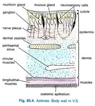
The body wall of Asterias consists of following tissue layers:
(i) Cuticle:
The body surface is clothed with a definite cuticle consisting of two layers, an outer thick homogeneous layer and an inner delicate layer.
(ii) Epidermis:
Just beneath the cuticle lies a layer of ciliated epithelium which extends over all the external appendages of body such as spines, pedicellariae, tube feet and gills, etc.
The epidermis is composed of a variety of cells such as ordinary flagellated or ciliated columnar cells, neurosensory cells, mucous gland cells or goblet cells having finely granular contents, muriform gland cells filled with coarse spherules and the pigment granules which provide characteristic external colouration to the animal.
(iii) Nervous Layer:
Beneath the epidermis lies a nervous layer, varying in thickness in different areas and penetrated by the attenuated bases of the epidermal cells on their elastic filaments.
(iv) Basement Membrane:
Just below the nervous layer lies a delicate basement membrane which separates the nervous layer and epidermis from the underlying dermis.
(v) Dermis:
The dermis is composed of fibrous connective tissue developed from the mesoderm. It is the thickest layer of body wall and has two regions outer and inner. The outer dermal region secretes and houses the endoskeletal ossicles and binds them together, while the inner dermal region contains numerous blood containing spaces called perihaemal spaces.
(vi) Muscular Layer:
The muscular layer consists of smooth muscle-fibres. It is differentiated into an outer circular muscle layer and inner longitudinal muscle layer. These muscle layers are on the whole weakly developed except in the aboral wall where stronger longitudinal bundles radiate from the centre of the disc along the mid-dorsal line of each arm, to bend the arms aborally.
(vii) Coelomic Epithelium:
The innermost layer of body wall lines the coelom and is composed of flagellated cuboidal cells of mesodermal origin. The innermost layer of body wall is called coelomic epithelium or peritoneum.
5. Endoskeleton of Asterias:
The rigidity of the body of Asterias is due to the presence of definite skeleton. In Asterias, the endoskeleton is unique in being mesodermal in origin instead of ectodermal as in other invertebrates. It consists of numerous calcareous ossicles. The ossicles are of various shapes and are bound together by connective tissue. They form a reticulate skeleton, leaving spaces for the emergence of groups of papulae.
The ossicles have irregular arrangement on the aboral surface but have definite and regular arrangement on the oral surface. On the oral surface, they are regularly around the mouth and in the ambulacral groove. Five plate-like ossicles called oral ossicles remain arranged around the mouth. Each ambulacral groove is supported by double rows of large, transversely placed opposite rod-shaped ambulacral ossicles.
The ossicles of the two opposite rows are arranged like an inverted V, their aboral ends meeting at the apex of the V, like the rafters supporting the roof of a shed and forming a conspicuous ambulacral ridge. The ambulacral ossicles do not bear any spines, tubercles or other external appendages. Because they are movably articulated in the ambulacral groove, they permit the opening or closing of the latter.
Further, each ambulacral ossicle has a notch on its outer as well as inner margin. The two notches of the adjacent ossicles together form an oval aperture, the ambulacral pore for the passage of tube-foot. The ambulacral pores are so arranged that they form two rows on each side of the ambulacral groove.
At its outer end, each ambulacral groove articulates with one admbulacral ossicle forming the edge of the groove and bearing two or three movable spines on small tubercles. Next to the adambulacral ossicle there are two rows of the ossicles called supra- and inframarignal ossicles.
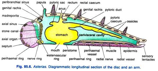
Section of an Arm:
The arm is covered all around by a thin two-layered cuticle, a ciliated epidermis and an underlying thick dermis which has many perihaemal spaces and ossicles.
Epidermis and dermis are thinned over the projecting spines, pedicellariae and papulae but they wear off from spines. The aboral side is a thick convex arch, and the oral side is like an inverted a, between the two limbs of the a is an ambulacral groove. The arm encloses a perivisceral coelom.
The aboral wall has a number of irregular, fenestrated ossicles which are calcareous, on some ossicles rest projecting spines.
From the aboral side dermal papulae project out, the coelom is continued into the papulae. Between the spines and attached to them are many pedicellariae. Each lateral margin of the arm has two large spines, they are a supra marginal spines and below it an inframarignal spine. Mid-dorsally the arm has a large cardinal spine.
On the oral surface the ambulacral groove is supported by two elongated ambulacral ossicles meeting at the summit of the groove, at each end of the ambulacral groove is an adambulacral ossicles and spine. The ambulacral ossicles form two columns in the oral surface of each arm and on each side there is a single column of adambulacrals.
The adambulacral spine can touch the substratum or bend inwards to protect the ambulacral groove. Above the ambulacral groove runs a radial canal which is joined on each side by a podial branch to two ampullae and one tube foot. Below the radial canal is a radial hyponeural sinus enclosing a radial haemal channel.
Muscles:
The median aboral side below the body wall has an apical longitudinal muscle which stretches the arm. Each pair of ambulacral ossicles has an upper and a lower transverse ambulacral muscle, the upper or superior transverse ambulacral muscle widens the ambulacral groove, and the lower or inferior transverse ambulacral muscle makes the ambulacral groove narrow.
Between the adjacent ambulacral ossicles of each side is a longitudinal ambulacral muscle which shortens the arm and the ambulacral groove. The outer end of each ambulacral ossicle is connected to the adambulacral ossicle by a lateral transverse ambulacral muscle which widens the ambulacral groove.
Nerves:
In the middle of the ambulacral groove is a radial nerve cord in the shape of a V. Above the radial nerve cord are two Lange’s nerves. Close to the outer end of each ambulacral ossicle is a marginal nerve. Each podium has a nerve ring.
Inside the perivisceral coelom of the arm is a pair of pyloric caeca, each suspended by two longitudinal mesenteries from the aboral surface. If the section passes through the base of the arm the perivisceral coelom has a pair of gonads attached to the body wall by their ducts.
6. Coelom in Asterias:
Asterias possesses a true and spacious coelom which is lined by a coelomic epithelium of ciliated cuboidal cells.
It consists of various compartments, viz.:
(i) A perivisceral coelom extending in central disc and rays and surrounding the visceral organs such as digestive tract and the gonads,
(ii) Coelom of water vascular system,
(iii) Axial sinus,
(iv) Perihaemal sinus and canals and,
(v) Genital sinuses, etc.
The coelom is filled with a colourless, alkaline coelomic fluid which contains various dissolved nutrients such as amino acids, fatty acids, glycerol and glucoses, etc. Besides nutrients, the coelomic fluid also contains two main types of phagocytic amoeboid corpuscles, the amoebocytes or coelomocytes; coelomocytes with ordinary slender pseudopodia and coelomocytes with petaloid pseudopodia.
The coelomic fluid, like the haemolymph of Arthropoda, bathes the tissue of the body and performs the function of circulatory system. It distributes the nutrients to various body cells and also performs the respiratory as well as excretory functions.
7. Digestive System of Asterias:
Alimentary Canal of Starfish:
In Asterias, the alimentary canal is tubular, straight, short and extends vertically along the oral-aboral axis in the central disc.
It comprises the following parts:
1. Mouth:
The mouth is the anterior most aperture of alimentary canal and it is situated in the centre of the peristomial membrane of the oral surface. It is provided with a sphincter muscle and radial fibres and is capable of great expansion and retraction. The mouth leads upward into the oesophagus.
2. Oesophagus:
The oesophagus is a very short, wide and vertical tube. It opens aborally in the stomach.
3. Stomach:
The stomach is a broad sac and fills the interior of the disc. It is typically divided by a horizontal constriction into a voluminous oral part, the cardiac stomach and a flattened aboral part, the pyloric stomach. The cardiac stomach has a muscular, highly folded wall bulged out to form five lobes, one opposite each arm.
The cardiac stomach is connected to the ambulacral ridge of each arm by ligaments of muscles and connective tissues called mesenteries or gastric ligaments which serve to anchor the cardiac stomach in place. During the feeding process, the cardiac stomach can be everted through the mouth by the contraction of muscles of body wall.
The retraction of cardiac stomach is brought about by five pairs of retractor muscles which arise from the lateral sides of the ambulacral ridge. The pyloric stomach is much smaller, flat and pentagonal sac. It communicates with the intestine.
4. Intestine:
The intestine is a short, narrow, five sided tube that runs straight upward to open out at the anus. It gives off two or three little hollow diverticula called intestinal or rectal caeca placed inter-radially. The intestinal caeca are brown in colour and each bears several short, irregularly-shaped diverticula. The actual function of intestinal caeca is well disputed. However, they are considered as excretory organs, because, they secrete a brown fluid.
5. Anus:
The intestine opens on the aboral surface by a posterior most aperture of the alimentary canal called anus. The anus is situated eccentrically on the aboral side of central disc.
Histology of Alimentary Canal:
Histologically the wall of the alimentary canal consists from within outwards of an enteric epithelium of columnar ciliated cells of endodermal origin, a sub-epidermal nervous layer, a connective tissue layer devoid of ossicles, a layer of muscles and an outermost layer of coelomic epithelium or visceral peritoneum. The thickness of the layers varies in different parts of the alimentary canal.
Digestive Glands:
To the pyloric stomach are attached ten, long brownish or greenish glandular appendages variously called pyloric caeca, digestive glands, branchial caeca, hepatic caeca, etc. There are two pyloric caeca in each arm, each suspended from the aboral wall of the arm by two longitudinal mesenteries that enclose between them a coelomic space continuous at its central end with the general coelom of the disc.
Each pyloric caecum consists of double series of hollow lobulated sacs that open into a central tube duct. The two ducts forming a pair of caeca of each arm unite to form a main pyloric duct that opens into the pyloric stomach at one of its angles.
Histology of Digestive Gland:
Histologically the pyloric caeca are complex structures and are lined by ciliated columnar epithelium which is composed of four types of epithelial cells.
The current producer cells bear longer flagella and maintain a steady circulation of the fluids and digested food in the cavities of the caeca; the mucous cells produce mucus; the secretory or granular cells secrete digestive enzymes to convert proteins into peptones, starch into monosaccharide sugars and fats into fatty acids and glycerol, and the storage cells store reserve food such as lipids, glycogen and polysaccharide-protein complex.
The pyloric caeca function like pancreas of vertebrates.
Physiology of Digestive System:
Food:
Asterias is a carnivorous animal and feed voraciously on worms, crustaceans, snails, bivalves, small-sized starfishes, echinoderms and fishes. Sometimes, Asterias also feed on dead animals and under hazardous conditions may live without food for several months.
Ingestion and Digestion:
The mode of feeding or ingestion in Asterias is of most unusual type. It swallows small-sized animals directly through the mouth. The prey is held by the tube feet and cardiac stomach is everted and wrapped round it. The enzymatic secretions of pyloric caeca poured out on to the prey and when digestion is completed, stomach is withdrawn along with the digested food.
To feed shelled molluscs (bivalves), Asterias adopts another interesting technique. It creeps over the clam and holding it with tube feet, orients it to bring the free margins of the shell close to its mouth. It now arches its body, assuming a characteristic humped or umbrella-like posture. The more proximal tube feet firmly grip both valves of the bivalve’s shell, while the more distal ones are attached to the substratum.
The cardiac stomach is now everted through the mouth by the concentration of body wall and pressure of coelomic fluid.
The tube feet gripping the valves of the clam’s shell exert a steady pull as the muscles in the arms contract. But, because the muscles of clam cannot maintain a continuous state of contraction for a long time and sooner or later, the adductors of the mussel become fatigued or exhausted and finally relax, so that the shell opens.
Asterias, now, inserts its already everted cardiac stomach into the mantle cavity of the clam and pours out its proteolytic enzymes.
The enzymes digest the visceral organs of clam, they; thus, convert the proteins of visceral organs into peptones and amino acids; polysaccharide carbohydrates and lipid contents into fatty acids and glycerol. When the digestion is partially completed the sea star withdraws its stomach along with the digested food by means of its retractor muscles and moves on leaving behind the shell of the victim.
Remaining digestion of food substances occurs in cardiac stomach from which the digested food diffuses to coelomic fluid for body wide distribution and to pyloric caeca where it may be distributed to arms or stored in storage cells of epithelium of pyloric caeca. The undigested food is either egested out directly from the mouth or pass through intestine and egested out through the anus.
8. Water Vascular System of Asterias:
The water vascular system is a modified part of coelom and it consists of a system of sea- water filled canals having certain corpuscles. It plays most vital role in the locomotion of the animal and comprises madreporite, stone canal, ring canal, radial canal, Tiedeman’s bodies, lateral canals, and tube feet.
(i) Madreporite:
As already stated, the madreporite is a rounded calcareous plate occurring on the aboral surface of the central disc in inter-radial position. Its surface bears a number of radiating, narrow, straight or wavy grooves or furrows. Each furrow contains many minute pores at its bottom.
Each pore leads into a very short, fine, tubular pore canal which passes inward in the substance of the madreporite. There may be about 200 pores and pore-canals. The pore-canals unite to form the collecting canals which open into an ampulla beneath the madreporite.
(ii) Stone Canal:
The ampulla opens into a S-shaped stone canal. The stone canal extends downwards (orally) and opens into a ring canal, around the mouth. The walls of stone canal are supported by a series of calcareous rings. The lumen of stone canal is lined by very tall flagellated cells.
In embryonic stages and in young Asterias, the stone canal remains a simple tube but in adult Asterias, lumen of stone canal possesses a prominent ridge with two spirally rolled lamellae which by branching become more complicated in structure. During its course, the stone canal is en-sheathed by a wide, thin-walled tubular coelomic sac, called axial sinus.
(iii) Ring Canal:
The ring canal or water ring is located to the inner side of the peristomial ring of ossicles and directly above (aboral) to the hypo neural ring sinus. It is wide and pentagonal or five sided.
(iv) Tiedemann’s Bodies:
The ring canal gives out inter-radially nine small, yellowish, irregular or rounded glandular bodies called racemose or Tiedemann’s bodies, from its inner margins. The Tiedemann’s bodies rest upon the peristomial ring of ossicles. The actual function of Tiedemann’s bodies is still unknown, however, they are supposed to be lymphatic glands to manufacture the amoebocytes of the water vascular system.
(v) Polian Vesicles:
The ring canal gives off on its inner side in the inter-radial position one, two or four, little, pear-shaped, thin-walled, contractile bladders or reservoirs with long necks called polian vesicles. They are supposed to regulate pressure inside ambulacral system and to manufacture amoeboid cells of ambulacral system.
(vi) Radial Canal:
From its outer surface the ring canal gives off a radial water canal into each arm that runs throughout the length of the arm and terminates as the lumen of terminal tentacle. In the arm the radial water canal runs immediately to the oral side of the ambulacral muscles.
(vii) Lateral Canals:
In each arm, the radial canal gives out two series of short, narrow, transverse branches called lateral or podial canals. Each lateral canal is attached to the base of a tube foot and is provided with a valve to prevent backward flow of fluid into the radial canal.
(viii) Tube Feet:
As already mentioned, there are four rows of tube feet in each ambulacral groove. A tube foot or podium is a hollow, elastic, thin-walled, closed cylinder or sac-like structure having an upper sac-like ampulla, a middle tubular podium and a lower disc-like sucker.
The ampulla lies within the arm, projecting into the coelom above the ambulacral pore which is a gap between the adjacent ambulacral ossicles for the passage of the podium. The tube feet are chief locomotory and respiratory organs of Asterias.
9. Locomotion of Asterias:
Asterias lacks in head or anterior end, therefore, capable to move in any direction according to its desire. It can move on horizontal as well as on vertical surfaces by the help of tube feet.
Locomotion on a Horizontal Surface:
When an Asterias desires to move on a horizontal surface in a given direction, the arm or arms pointing in that direction is lifted.
The ampullae of raised arm contract, the valve in the lateral canals close and the water of the ampullae is forced into the podia. The podia of the tube feet become extended, elongated and enlarged in the general direction of movement due to the hydrostatic pressure produced by influx of water into them.
Subsequently, the terminal suckers of the tube feet become attached to the substratum and their central parts are withdrawn to form suction cups. Due to the vacuum so produced, the suckers acquire a firm grip over the substratum. Mucus secreted by the tips of the tube feet further aids in attachment.
The tube feet now pivot forward on their attached suckers, assuming vertical position and thereby pushing the body forwards. The longitudinal muscles of the podia now contract and this forces their fluid back into the ampullae and releases their suckers. The ampullae then contract again and whole sequence of events is repeated.
Locomotion on a Vertical Surface:
In climbing a vertical surface, the tube feet pull the body forward. By the alternate contraction and expansion of tube feet and by adherence of suckers of tube feet on surface Asterias climbs on the vertical surface.
Asterias employs its tube feet, only when, it moves on hard rocky substratum. But, on soft mud or sand (substratum) the suckers of tube feet become useless, therefore, on such soft surfaces the animal literally walks on its extended tube feet which now act like small legs. Besides locomotion, tube feet serves many other functions such as clinging of animal body to substratum, tactile and respiratory function.
10. Circulatory System of Asterias:
The so-called circulatory system includes following two systems:
1. Perihaemal system
2. Haemal system
1. Perihaemal System:
The perihaemal system, like the water vascular system, is derived from the coelom and is composed of many tubular coelomic sinuses such as axial sinus, aboral ring sinus, genital sinuses, radial perihaemal sinuses, marginal sinuses and peribranchial sinuses.
(i) Axial Sinuses:
The axial sinus is a thin-walled, vertical, wide tubular coelomic cavity enclosing the stone canal and the axial gland. The three forming a well developed axial complex.
(ii) Aboral Ring Sinus:
The aboral ring sinus is a tubular, pentagonal channel or sinus around the intestine, lying just inside the aboral wall of the central disc. It communicates with the axial sinuses.
(iii) Genital Sinuses:
The aboral ring sinus gives off five pairs of genital branches, one pair in each arm. The genital sinuses surround the gonads.
(iv) Oral Ring Sinus:
At its oral end, the axial sinus opens into the inner division of a circular channel, the oral, peribuccal, perihaemal, or hypo neural ring sinus which runs around the mouth. It is a large tubular sinus and is divisible into an inner narrow and an outer wide ring by an oblique circular septum called haemal strand.
(v) Radial Perihaemal Sinuses:
The outer division of ring sinus gives out five radial hypo neural or perihaemal sinuses, one of which extends through each arm between the radial nerve and the radial water canal. Like oral ring sinus, each radial sinus is also divided longitudinally into two by a vertical partition or septum, continuous with the haemal strand. The radial perihaemal sinuses also give out fine channels into the tube feet.
(vi) Marginal Sinuses:
In each arm, two longitudinal marginal sinuses run longitudinally on each side just aboral to the marginal nerve cord. The fine lateral channels connect the marginal channels with the radial perihaemal sinuses.
(vii) Peribranchial Sinuses:
The sinuses occurring as circular spaces around the basal parts of papulae or gills are called peribranchial sinuses.
2. Haemal System:
The haemal or blood lacunar system of Asterias is reduced and is of open type like the haemocoel of Arthropoda and Mollusca. It includes inter-communicating spaces having no coelomic epithelium and are derived embryo-logically from the blastocoel. The haemal system is filled with coelomic fluid containing coelomocytes and is enclosed in the coelomic spaces of perihaemal system.
The main haemal sinuses are as follows:
(i) Oral Haemal Ring:
It is the circular haemal sinus, located around the mouth just below the– ring canal of the water vascular system. Oral haemal ring is a fine channel or a ring of lacunar tissue which runs in the septum dividing the hyponeural sinus. The oral haemal ring is connected with aboral haemal ring through axial gland.
(ii) Radial Haemal Sinuses or Strands:
These arise radially from the oral haemal ring and one extends into each arm, along the floor of the ambulacral groove just below the radial canal of the water vascular system. The radial haemal sinuses also give off branches into the podia.
(iii) Axial Complex:
The perihaemal and haemal systems of Asterias are intimately connected by a complicated structure called axial complex. The axial complex comprises the following three parts a thin-walled, tubular coelomic cavity called axial sinus containing the stone canal of water vascular system and axial gland, both are closely attached with the wall is of axial sinus by the mesenteries.
(iv) Axial Gland:
This is the principal part of the haemal system. The axial gland is an elongated, fusiform, brownish or purple coloured spongy body. It is covered externally by coelomic epithelium and is called variously as heart, ovoid gland, dorsal organ, septal organ, brown gland, etc. The axial gland is connected with the oral and aboral haemal sinuses at its oral and aboral ends respectively.
At its oral end the axial gland becomes thin and terminates in the septum that subdivides the hypo neural ring sinus. At its aboral end, the axial gland has an aboral extension or terminal process called head process which is lodged in a separate, closed contractile coelomic sac called dorsal sac.
The dorsal sac is situated below the madreporite, close to the ampulla of the stone canal, but has no communication with the ampulla. A pair of gastric tufts also arises from the haemal sinuses in the wall of the cardiac stomach and opens into the axial gland near its aboral end. Digested food from the stomach passes into the haemal circulation through the gastric tufts.
Histologically, the axial gland has an external lining of peritoneum and its interior is filled by connective tissue outlining numerous spaces containing irregularly arranged cells of the nature of coelomocytes. The axial gland has an intimate relation with the circulation of blood in perihaemal and haemal channels.
(v) Aboral Haemal Ring:
It is a pentagonal ring canal lying beneath the aboral surface of the central disc. From the aboral haemal ring or canal extend five pairs of genital haemal strands to the gonads.
Function:
The haemal system acts as a pathway for the distribution of food substances carried by the coelomocytes. The flow of fluid within it is maintained by the contractile activity of the dorsal sac. The axial gland acts as a genital stolon, producing sex-cells, which reach the gonads through the aboral haemal ring and its branches.
11. Respiratory and Excretory System of Asterias:
The respiratory organs of Asterias are gills or papulae and tube feet. The papulae are the chief respiratory organs. They are simple, contractile, transparent, hollow outgrowths of body wall on the aboral surface having ciliated epithelium at outer and inner surfaces. They are derived from coelom and their lumen remains in direct communication with coelom.
An exchange of O2 and CO2 takes place between sea water and body fluid of gills. The cilia of epithelial cells have vital role in movement of coelomic fluid and in creating respiratory water currents in sea water. The other thin- walled, richly vascularized and moist body parts also act as respiratory organs.
Excretory System of Asterias:
Asterias lacks well specialised excretory organs. The nitrogenous metabolic excretory waste usually contains ammonium compounds. They pass from various tissues into the coelomic fluid and from there they diffuse through the thin walls of the rectal caeca, tube feet and gills. The coelomocytes have significant role in the excretion of excretory wastes from the coelom.
12. Nervous System of Asterias:
The nervous system of Asterias is of simple and primitive type. It is composed of nerve fibres and nerve nets which are closely associated with the epidermis. The nervous system comprises the following four units placed at different levels in the disc and arms.
(i) Oral or Ectoneural or Epidermal Nervous System:
The oral nervous system is situated just beneath the epidermis. It is the main part of the sensory nervous system and sensory in nature.
It comprises the following parts:
(a) Nerve ring:
The nerve ring is pentagonal in shape and is circum-oral, i.e., occurs around the mouth in the peristomial membrane. It supplies nerve fibres to the peristomial membrane and the oesophagus and at each radius gives off a radial nerve.
(b) Radial nerve:
The nerve ring gives off five radial nerves, each of which runs throughout the length of the arm in the bottom of the ambulacral groove. Each radial nerve terminates as a sensory cushion on the aboral side of the terminal tentacle.
A cross section of an arm shows that the radial nerve is a thick V-shaped mass continuous on its outer side with the epidermis and separated on its inner side from the hypo neural sinus only by a thin dermis and the coelomic epithelium. The radial nerve consists of fibrillae arranged in layers and interspersed with bipolar and multipolar ganglion cells.
(c) Sub-epidermal nerve complex:
The sub-epidermal nerve complex is an extensive network of nerve cells and nerve fibres beneath the epidermis all over the body surface including the gills and pedicellariae, etc. It is connected with radial nerve cords by nerve fibres.
The sub-epidermal nerve-plexus is thickened into a cord which forms:
(i) Two marginal nerves each of which extends throughout the length of an arm on each side and gives off a longitudinal series of lateral motor nerves which supply the ossicles, muscles, coelomic epithelium of that area and
(ii) A nerve ring in the suckers of each tube foot.
The ectoneural nervous system acts as the central nervous system of Asterias. It has sensory and motor neurons.
(ii) Deep or Hypo Neural Nervous System:
The hypo neural nervous system occurs in the form of a nervous layer in the lateral part of the oral wall of the hypo neural sinus, beneath the coelomic epithelium lining the sinus. This nervous layer is called Lange’s nerve. It is separated from the lateral part of the radial nerve only by a thin layer of dermal connective tissue.
Lange’s nerve gives off a series of nerves along the arm into the adjacent lower transverse muscle extending between the ambulacral ossicles in the roof of the hypo neural sinus. Lange’s nerve continues to the peristomial region, where it forms five inter-radial thickenings in the floor of the ring sinus that lies aboral to the main nerve ring.
(iii) Aboral or Coelomic Nervous System:
The aboral or coelomic nervous system is situated just outside the parietal peritoneum on the aboral side. It consists of a nerve ring in the central disc and a nerve in each arm. This system has connection with marginal nerves by nerve fibres. It innervates the body muscles of aboral side and is motor in function.
(iv) Visceral Nervous System:
The visceral nervous system occurs in wall of the gut just outside the enteric epithelium. It innervates the muscles of alimentary canal and is connected with the visceral receptors.
13. Sense Organs of Asterias:
Asterias possesses a few primitive sense organs which are as follows:
(i) Eyes:
The eyes are the most significant sensory organs of Asterias. They are simple, pigmented and occur at the base of terminal tentacles. On the oral surface, at the base of each terminal tentacle occur optic cushions which are composed of the thick epidermis with many photoreceptors or pigmented cup ocelli.
Each ocellus is a cup-shaped or funnel-like pocket of ectoderm. It is covered externally by the cuticle beneath which is found in many species a lens formed by the epidermis. The wall of cup consists of epidermal cells, altered into a shorter,- stouter shape and filled distally with red pigment granules and of retinal cells, disposed between the pigment cells.
The retinal cells are elongated cells with a distal bulbous enlargement projecting into the cavity of the cup and a proximal fibre passing into the underlying radial nerve. The number of ocelli in one optic cushion or eyes ranges from 80 to 200 in different species. A transparent gelatinous tissue fills the cavity of ocellus. The ocelli are light-perceiving organs which can detect changes in light intensity.
(ii) Terminal Tentacles:
The terminal tentacles have sensory cells which are tactile and also sensitive to food and other chemical stimuli.
(iii) Neurosensory Cells:
The entire body surface or epidermis of Asterias is traversed by many neurosensory cells serving as both tango- and chemoreceptors. The neurosensory cells are slender cells with a fusiform body containing the nucleus, a distal thread-like process reaching to the cuticle, and a proximal fibre entering the sub-epidermal nerve plexus.
They are especially numerous in the suckers of the podia, at the base of spines and pedicellariae and terminal tentacles.
14. Reproductive System of Asterias:
Most species of Asterias are unisexual or dioecious, i.e., sexes are separate except a few species such as Asterias rubens which is hermaphrodite. There is no marked sexual dimorphism, however, during breeding season some sort of colour difference between both the sexes may occur.
The reproductive organs of Asterias are of primitive type and lack copulatory organs, accessory glands, receptacles for storing ova and reservoirs for storing mature sperms. There are only gonads which act as reproductive organs.
Gonads:
The male gonads are testes and female gonads are ovaries. Each sexually mature male or female individual contains five pairs of testes or ovaries, one pair is lying free laterally in the proximal part of each arm between the pyloric caeca and the ampullae.
The testes and ovaries are morphologically similar. Each gonad appears as an elongated feathery tuft or tuft of tubules or bunch of grapes, whose size varies greatly according to the proximity of spawning time.
At maturity the gonads occupy the entire perivisceral space. The proximal end of each gonad is attached to aboral body wall near the inter-brachial septum by a very short gonoduct which is ciliated and opens laterally through a small gonopore on the aboral surface almost at the angle of two adjacent arms.
Each gonad is enclosed in a genital sac of coelomic nature with a wall of muscle and connective tissue fibres, covered externally with peritoneum. This genital sac is the outgrowth of the genital or aboral coelomic sinus. The gonad proper is lined by germinal epithelium, containing the germ cells.
The mature sperms and ova are discharged by male and female Asterias respectively in sea water. The release of sex cells from the gonads is regulated by neuro-hormonal secretion of radial nerve.
15. Life History and Development of Asterias:
(i) Fertilisation:
The most species of Asterias have only one breeding season in a year. During breeding season, both types of mature sexes shed their sex cells in the sea and union of male and female sex cells or gametes (sperms and ova) occurs in sea water. Thus, fertilisation in Asterias is external.
(ii) Embryogeny:
The embryological development of Asterias is indirect and includes various larval stages. The fertilised egg or zygote is spherical, half millimeter in diameter and contains little amount of yolk. The cleavage is holoblastic and equal and it converts the unicellular zygote into a single layered, hollow, ciliated and spherical structure called coeloblastula.
The coeloblastula possesses a fluid-filled central space, called blastocoel and it swims about freely. The blastula undergoes embolic invagination and becomes two layered cup-like gastrula. The gastrulation involves the inward pushing of blastomeres of one side. The in-pushing encloses a cavity called archenteron and it occupies the larger part of blastocoel which ultimately becomes obliterated.
This embryonic stage is called gastrula and it has an outer ectodermal and inner endodermal germinal layers. The archenteron or gastrocoel communicates to the exterior by a wide aperture called blastopore. The blastopore changes its relative position with the elongation of gastrula and becomes the anal opening of the larva. Two more openings appear on the surface of the larva.
On the ventral side, a tubular in-growth of ectoderm forms the larval mouth or stomodaeum. Another opening occurs on the dorsal side as the dorsal pore. The cilia of general surface of gastrula degenerate and certain definite ciliary band appears. The mesoderm is formed from two sources.
During the gastrular invagination, the advancing tip of archenteron (endoderm) buds off certain mesenchyme cells into the blastocoel. The growing archenteron is differentiated into a narrow proximal part and wide terminal part.
The narrow proximal part communicates to the exterior by the blastopore and in later stages forms the stomach, and intestine, while the wide terminal part of completed archenteron expands and cuts off on each side into a coelomic pouch, the hydroenterocoel.
These take up their position to the right and the left sides of the archenteron and develop into coelomic pouches. The latter give rise to coelom, its mesodermal lining and water vascular system. The embryo at this stage becomes a free-swimming larva.
(iii) Larval Development:
The larval development of Asterias includes the following larval stages:
Bipinnaria Larva:
The bipinnaria larva develops from the zygote in about one week. It is a bilaterally symmetrical larva which possesses a preoral and a postoral ciliated band, and a preoral lobe with preoral loop of ciliated band. The various projections emerging out of its body correspond to the arms. Inside the body appears the coelomic apparatus and the alimentary canal.
The bipinnaria larva feeds on diatoms, etc., by creating food-bearing currents by ciliary tracts in the stomodael wall. It swims freely by forwarding its anterior end, with a clockwise rotation, after some time the bipinnaria larva transforms into the next larval stage, the brachiolaria larva.
Brachiolaria Larva:
In the brachiolaria larva the side-lobes of bipinnaria increase in length to become long, slender and ciliated larval arms. The larval arms move and contract. The preoral arms also give out processes called the brachiolar arms. The arms of brachiolaria larva have coelomic prolongations and possess tips of adhesive cells.
The bases of these arms surround the elevated, adhesive, glandular area performing the function of a sucker or fixation disc by which the larva becomes attached at the time of metamorphosis.
(iv) Metamorphosis:
In about 6 or 7 weeks, the brachiolaria larva settles on the bottom or on some solid object and is fixed with that by its adhesive arms. Now the bilaterally symmetrical larva metamorphoses into a radially symmetrical adult. The larval mouth and anus close. A new mouth is formed on the left side of the larva and a new anus is developed on the right side.
The left and right side of the larva, thus, subsequently differentiated into oral and aboral surfaces of the adult. Five lobes called arm rudiments grow out around oral-aboral axis. In later stages, the skeletal elements appear on the arm rudiments and the radial canals grow into them.
In each arm two pairs of outgrowths from the coelom form the first tube feet and serve for attachment. Further complex re-organisational changes result in the formation of adult Asterias. The newly detached rudiment of the body of sea star is less than 1 mm with short stubby arms.
16. Regeneration and Autotomy of Asterias:
Asterias possesses considerable power of regeneration. It is capable to regenerate its any lost part of body at any time. Moreover, if an arm is injured or held up, Asterias usually casts it off near the base at the fourth or fifth ambulacral ossicle. This is called autotomy.
The opening left in the side of the central disc by the broken off arm is immediately closed by the contraction of the adjacent body wall musculature for the protection of internal body organs and regeneration of new arm starts at that place.
A disc deprived of all its arms regenerates. In Asterina vulgaris, a single arm with a portion of disc regenerates an entire animal. But in Linckia, an arm totally devoid of disc can also regenerate complete animal (Fig. 85.15). Specimens with small regenerating arms at the base of the large original arm are popularly called comets.
