In this article we will discuss about Sponges:- 1. History of Sponges 2. Definition of Sponges 3. Origin 4. General Characters 5. Classification 6. General Organisation 7. Canal System 8. Skeleton 9. Reproduction 10. Development 11. Animal Nature 12. Affinities 13. Economic Importance.
Contents:
- History of Sponges
- Definition of Sponges
- Origin of Sponges
- General Characters of Sponges
- Classification of Sponges
- General Organisation of Sponges
- Canal System in Sponges
- Skeleton in Sponges
- Reproduction in Sponges
- Development of Sponges
- Animal Nature of Sponges
- Affinities of Sponges
- Economic Importance of Sponges
Contents
- 1. History of Sponges:
- 2. Definition of Sponges:
- 3. Origin of Sponges:
- 4. General Characters of Sponges:
- 5. Classification of Sponges:
- 6. General Organisation of Sponges:
- 7. Canal System in Sponges:
- 8. Skeleton in Sponges:
- 9. Reproduction in Sponges:
- 10. Development of Sponges:
- 11. Animal Nature of Sponges:
- 12. Affinities of Sponges:
- 13. Economic Importance of Sponges:
1. History of Sponges:
The name Porifera (L., porus = pore; ferro = to bear) comes from (1836). The nature of sponges was debated until well into the nineteenth century, although evidence of their animal nature was given in 1765 by who saw the water currents and movements of the oscula. As a result Linnaeus, Lamarck, and classified the sponges under zoophytes or polyps and regarded them as allied to anthozoan coelenterates.
Although (1816) separated the sponges in a group Spongiaria allied to Protozoa. The morphology and physiology of sponges were first adequately understood by who created in 1836 the name Porifera for the group by which it is now generally known, iuxle (1875) and Sollas (1884) proposed the complete separation of sponges from other Metazoa on the grounds of many peculiarities.
Sponges are now recognised as constituting a separate isolated branch of the Metazoa named Parazoa, after Sollas.
Porifera include the sponges which are most primitive of multicellular animals, they are sessile, plant-like animals, they are fixed to some submerged solid rock or shell and are incapable of any movement. The Porifera are exclusively marine except for a single family of freshwater species.
Their shape may be cylindrical, branching,vase-like or globular, some are dull in colour but most are brightly coloured, they have red, orange, purple, green or yellow colour. The body is perforated by pores and canals but there are no organs, such as mouth or nervous system. Though sponges are multicellular animals their cells do not form organised tissues. They usually have an endoskeleton of separate spicules.
Digestion takes place within the cells. Because of their endoskeleton and obnoxious ferments they are generally not eaten by animals. Sponges are cultivated for commercial purposes.
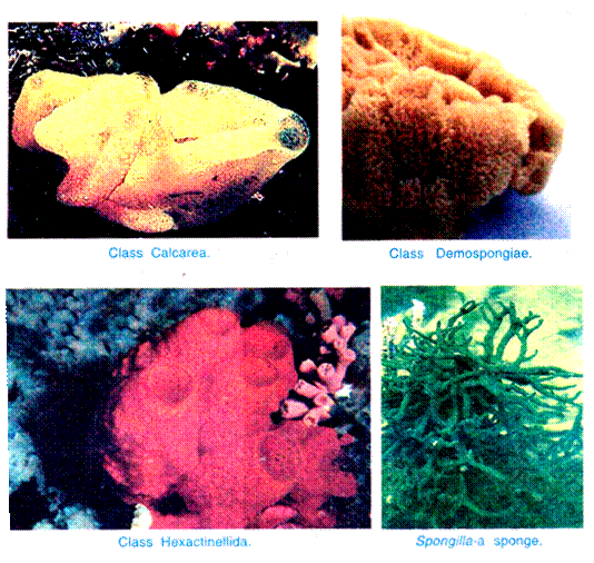
Approximately 10,000 species of sponges are known at present, and the phylum is divided into three classes, viz., Calcarea or Calcispongiae, Hexactinellida or Hyalospongiae, and Demospongiae and about twelve orders chiefly on the type of skeleton.
2. Definition of Sponges:
The Porifera may be defined as “asymmetrical or radially symmetrical multicellular organisms with cellular grade of organisation without well-defined tissues and organs; exclusively aquatic; mostly marine, sedentary, solitary or colonial animals with body perforated by pores, canals and chambers through which water flows; with one or more internal cavities lined with choanocytes; and with characteristic skeleton made of calcareous spicules, siliceous spicules or horny fibres of spongin”.
3. Origin of Sponges:
There is a great controversy regarding the origin of Porifera. However, they look more nearer to Protozoa though they differ from them in being multicellular. Porifera probably originated from flagellated protozoan like Proterospongia, a colonial flagellate. The colony of Proterospongia has collared and flagellated cells embedded in a gelatinous matrix having amoeboid cells.
These cells are the choanocytes and amoebocytes of sponges. Even then, the origin of sponges is believed to be uncertain. Conclusively it can be said that, sponges diverged from metazoan evolutionary line in the very early stage and, therefore, occupy a separate status.
4. General Characters of Sponges:
1. Porifera are all aquatic, mostly marine except one family Spongillidae which lives in freshwater.
2. They are sessile and sedentary and grow like plants.
3. Body shape is vase or cylinder-like, asymmetrical or radially symmetrical.
4. The body surface is perforated by numerous pores, the ostia through which the water enters the body and one or more large openings, the oscula by which the water passes out.
5. Multicellular body consisting of outer ectoderm and inner endoderm with an intermediate layer of mesenchyme, therefore, diploblastic animals.
6. The interior space of the body is either hollow or permeated by numerous canals lined with choanocytes. The interior space of sponge body is called spongocoel.
7. Characteristic skeleton consisting of either fine flexible spongin fibres, siliceous spicules or calcareous spicules.
8. Mouth absent, digestion intracellular; excretory and respiratory organs absent.
9. The nervous and sensory cells are probably not differentiated.
10. The sponges are monoecious; reproduction both by asexual and sexual methods.
11. Asexual reproduction occurs by buds and gemmules.
12. The sponges possess high power of regeneration.
13. Sexual reproduction occurs by ova and sperms; fertilisation is internal but cross- fertilisation occurs as a rule.
14. Cleavage holoblastic, development indirect through a free-swimming ciliated larva called amphiblastula or parenchymula.
15. The organisation of sponges has been grouped into three main types, viz., ascon type, sycon type and leuconoid type due to simplicity in some forms and complexity in others.
5. Classification of Sponges:
The classification of Porifera is based chiefly on types of skeleton found in them. This phylum has been classified variously but the classification suggested by Hyman (1940) and Burton (1967) are of considerable importance. However, classification of Porifera followed here is based on Storer and Usinger (1971) which appears to be a modified form of Hyman’s classification.
Class I. Calcarea (L., Calx = Lime) or Calcispongiae:
(L., calx = lime,Gr., spongos = sponge):
1. They have a skeleton of separate calcareous spicules which are monaxon or tetraxon; tetraxon spicules lose one ray to become triadiate.
2. They are solitary or colonial; body shape vase-like or cylindrical.
3. They may show asconoid, syconoid, or leuconoid structure.
4. They are dull-coloured sponges less than 15 cm in size.
5. They occur in shallow waters in all oceans.
Order 1. Homocoela:
1. Asconoid sponges with radially symmetrical cylindrical body.
2. Body wall is thin and not folded, spongocoel is lined by choanocytes.
Examples:
Leucosolenia, Clathrina.
Order 2. Heterocoela:
1. Syconoid or leuconoid sponges having vase- shaped body.
2. The body wall is thick and folded, choanocytes line only radial canals.
3. Spongocoel is lined by flattened endoderm cells.
Examples:
Sycon or Scypha, Grantia.
Class. II. Hexactinellida or Hyalospongiae:
(Gr., hyalos = glassy; spongos = sponge):
1. They are called glass sponges; skeleton is of siliceous spicules which are triaxon with six rays. In some the spicules are fused to form a lattice-like skeleton.
2. There is no epidermal epithelium.
3. Choanocytes line finger-shaped chambers.
4. They are cylindrical or funnel-shaped and are found in deep tropical seas, they grow up to one metre.
Order 1. Hexasterophora:
1. Spicules are hexasters, i.e., star-like in shape.
2. Radial canals or flagellated chambers are simple.
3. They are not attached by root tufts but commonly attached to a hard object.
Examples:
Euplectella, Farnera.
Order 2. Amphidiscophora:
1. Spicules are amphidiscs. No hexasters.
2. They are attached to the substratum by root tufts.
Examples:
Hyalonema, Pheronema.
Class III. Demospongiae (Gr., dermos = frame; spongos = sponge):
1. It contains the largest number of sponge species. Large-sized, solitary or colonial.
2. The skeleton may be of spongin fibres or of spongin fibres with siliceous spicules or there may be no skeleton.
3. Spicules are never six-rayed, they are monaxon or tetraxon and are differentiated into large megascleres and small microscleres.
4. Body shape is irregular and the canal system is of leucon type.
5. Generally marine, few freshwater forms.
Subclass I. Tetractinellida:
1. Sponges are mostly solid and simple rounded cushion-like flattened in shape usually without branches.
2. Skeleton comprised mainly of tetraxon siliceous spicules but absent in order Myxospongida.
3. Canal system is leuconoid type. Shallow water form.
Order 1. Myxospongida:
1. Simple structure.
2. Skeleton absent.
Examples:
Oscarella, Halisarca.
Order 2. Carnosa:
1. Simple structure.
2. Spicules are not differentiated into megascleres and microscleres.
3. Asters may be present.
Example:
Plakina.
Order 3. Choristida:
1. Spicules are differentiated into megascleres and microscleres.
Examples:
Geodia, Thenea.
Subclass II. Monaxonida:
1. Monaxonids occur in variety of shapes from rounded mass to branching type or elongated or stalked with funnel or fan-shaped.
2. Skeleton consists of monaxon spicules with or without spongin.
3. Spicules are distinguished into megascleres and microscleres.
4. They are found in abundance throughout the world.
5. Shallow and deep water forms.
Order 1. Hadromerida:
1. Monaxon megascleres in the form of tylostyles.
2. Microscleres when present in the form of asters.
3. Spongin fibres are absent.
Examples:
Cliona, Tethya.
Order 2. Halichondrida:
1. Monaxon megascleres are often of two types, viz., monactines and diactines.
2. Microscleres are absent. Spongin fibres present but scanty.
Example:
Halichondria.
Order 3. Poecilosclerida:
1. Monaxon megascleres are of two types, one type in the ectoderm and another type in the choanocyte layer.
2. Microscleres are typically chelas, sigmas and toxas.
Example:
Cladorhiza.
Order 4. Haplosclerida:
1. Monaxon megascleres are of only one type, viz., diactinal.
2. Microscleres are absent.
3. Spongin fibers are generally present.
Examples:
Chalina, Pachychalina, Spongilla
Subclass III. Keratosa:
1. Body is rounded and massive with a number of conspicuous oscula.
2. Skeleton composed of network of spongin fibres only.
3. Siliceous spicules are absent.
4. They are also known as horny sponges found in shallow and warm waters of tropical and subtropical regions.
Examples:
Euspongia, Hippospongia.
6. General Organisation of Sponges:
The general organisation of sponges varies considerably. The sponges are cylindrical like Leucosolenia, vase-shaped like Scypha and Grantia, tree-like (e.g., Microciona), finger-like (e.g., Haliclona), leaf-like (e.g., Phyllospongia), cushion-shaped like Euspongia, rope-like (e.g., Hyalonema), bowl-shaped like Pheronema, etc. Some sponges are solitary, while others are colonial.
The sponges are mostly attached forms; they are found attached to stones, shells, sticks, sea weeds, etc.; some are boring sponges like Cliona. Usually, the sponge body is asymmetrical but few forms exhibit radial symmetry. The size varies from few mm to massive having 1 or 2 metres diameter.
The body colouration of sponges also varies greatly; they are mostly white or grey but yellow, brown, purple, orange, red and green coloured species are also reported. The green colour of the sponge is usually due to the presence of a symbiotic algae, Zoo chlorella, in them.
7. Canal System in Sponges:
All the cavities of the body traversed by the currents of water, which nourish the sponge from the time it enters by the pores until it passes out by the osculum, are collectively termed canal system. In Olynthus, canal system is seen in its simplest type.
In other forms it may attain a high degree of complexity, but its general evolution can nevertheless be reduced to simple process of growth on the part of primitive Olynthus resulting in folding of the walls and accompanied by a restriction of the collared (choanocyte) cells to certain regions.
In the gradual and continuous process of differentiation, three distinct types of organisation can be distinguished which connected by numerous transitions may yet be considered as three styles of architecture, so to speak under which all existing forms may be classified. There are usually three types of canal system met within sponges, viz-, asconoid type, syconoid type and leuconoid type.
(i) Asconoid Type:
Asconoid type of canal system is the simplest of all the types. In this there is a radially symmetrical vase-like body consisting of a thin wall enclosing a large central cavity the spongocoel opening at the summit by the narrowed osculum.
The wall is composed of an outer and inner epithelium with a mesenchyme between. The outer or dermal epithelium here termed epidermis consists of a single layer of flat cells. The inner epithelium, lining the spongocoel, is composed of choanocytes. The mesenchyme contains skeletal spicules and several types of amoebocytes, all embedded in a gelatinous matrix.
The wall of the asconoid sponge is perforated by numerous microscopic apertures termed incurrent pores or ostia which extend from the external surface to the spongocoel. Each pore is intracellular, i.e., it is a canal through a tubular cell called a porocyte.
The water current impelled by the flagella of the choanocytes passes through the incurrent pores into the spongocoel and out through the osculum (water from exterior → incurrent pores → spongocoel → osculum → water out) furnishing in its passage food and oxygen and carrying away metabolic wastes. Asconoid type of canal system is found only in few sponges, e.g., Olynthus, Leucosolenia.
According to Hyman, the important features of the asconoid structure are the simple wall and the complete continuous lining epithelium of choanocytes, interrupted only by the inner ends of the porocytes. The asconoid type of sponge superficially resembles a typical gastrula.
2. Syconoid Type:
Syconoid type of canal system is the first stage above the asconoid type. It is formed by the out-pushing of the wall of an asconoid sponge at regular intervals into finger-like projections, called radial canals.
At first these radial canals are free projections and the outside water surrounds their whole length, for there are no definite incurrent channels. But in most syconoid sponges, the walls of the radial canals fuse in such a manner as to leave between them tubular spaces, the incurrent canals, which open to the exterior between the blind outer ends of the radial canals by apertures termed dermal ostia or dermal pores.
Since these incurrent canals represent the original outer surface of the asconoid sponge, they are necessarily lined by epidermis. Radial canals being the outpushings of the original spongocoel are necessarily lined by choanocytes and are, therefore, better called flagellated canals.
The interior of the syconoid sponge is hollow and forms a large spongocoel which is lined by the flat epithelium derived from epidermis. The openings of the radial canals into the spongocoel are termed internal ostia. The syconoid sponges retain the radial vase form of the asconoids and the spongocoel opens to the exterior by the single terminal osculum.
The wall between the incurrent and the radial canals, is pierced by numerous minute pores called prosopyles. The water current in syconoid sponges takes the following route dermal pores → incurrent canals → prosopyles → radial canals → internal ostia (apopyles) → spongocoeL → osculum → out.
The syconoid sponges differ from the asconoid type in two important particulars:
(i) In the thick folded walls containing alternating incurrent and radial canals and
(ii) In the breaking of the choanocyte layer, which no longer lines the whole interior but is limited to certain definite chambers (radial canals).
The syconoid structure occurs in two main stages. The first type illustrated in a few of the heterocoelous calcareous sponges, especially members of the genus Sycon. In the second stage, the epidermis and mesenchyme spread over the outer surface forming a thin or thick cortex often containing special cortical spicules. The epidermis becomes pierced by more definite pores than lead into narrowed incurrent canals.
3. Leuconoid Type:
As a result of further process of out folding of the choanocyte layer and thickening of body wall, the leuconoid type of canal system develops. The choanocyte layer of the radial canal of the syconoid stage evaginates into many small chambers, and these may repeat the process, so that clusters of small rounded or oval flagellated chambers replace the elongated chambers of the syconoid stage.
The choanocytes are limited to these chambers. Mesenchyme fills in the spaces around the flagellated chambers. The spongocoel is usually obliterated and the whole sponge becomes irregular in structure and indefinite in form. The interior of the sponge becomes permeated by many incurrent and ex-current canals join to form larger ex-current canals and spaces which lead to the oscula.
The surface is covered with epidermal epithelium and is pierced by many dermal pores (ostia) and oscula.
The dermal pores lead into incurrent canals that branch irregularly through the mesenchyme. The incurrent canals lead into the small rounded flagellated chambers by opening still termed prosopyles. The flagellated chambers open by apertures called apopyles into ex-current channels, and these unite to form larger and larger tubes, of which the largest lead to the oscula.
The main characteristics of the leuconoid type of canal system are the limitation of the choanocytes to small chambers, the great development of the mesenchyme, and the complexity of the incurrent and excurrent canals.
The course of water current is dermal ostia → incurrent canals → prosodus (if present) → prosopyles → flagellated chambers → apopyles → aphodus (if present) → excurrent canals → larger channels → oscula → out. The leucon type of canal system is very efficient and most sponges are built on the leuconoid plan and they attain a considerable size.
They are always irregular in structure but the flow of the current of the water is fairly rapid and efficient. The leuconoid type of canal system exhibits numerous variations but presents three stages of evolution, viz., eurypylous, aphodal, and diplodal.
(a) Eurypylous:
In the eurypylous leuconoid type of canal system, the flagellated chambers are wide and thimble-shaped, each opening directly into the excurrent canal by a wide aperture called apopyle and receive the water supply direct from the incurrent canal through the prosopyle.
The current of water takes the following route → dermal pores or ostia → subdermal spaces → incurrent canals → prosopyles → flagellated chambers → apopyles —> excurrent canals spongocoel → oscula → out. This type of canal system is found in Leucilla.
(b) Aphodal:
In the aphodal leuconoid type of canal system, the flagellated chambers are small and rounded. The opening of each flagellated chamber into the excurrent canal is drawn out into a narrow tube, usually not of great length, termed aphodus. The relation of the flagellated chambers to the incurrent canals remain as before.
The route of water current is as follows dermal pores or ostia → subdermal space → incurrent canals → prosopyles → flagellated chambers → aphodus → excurrent canals→ spongocoel → oscula → out. This type of canal system is found in Geodia and Stelleta.
(c) Diplodal:
In some cases there is also a narrow current tube, the prosodus between the incurrent canal and the flagellated chambers, such a condition is called diplodal. This type of canal system is found in Oscarella, Spongilla, etc.
The current of water takes the following route:
Dermal pores or ostia → subdermal spaces → incurrent canals → prosodus → flagellated chambers → aphodus → excurrent canals → spongocoel → oscula → out.
4. Rhagon Type:
In calcareous sponges, the leuconoid structure may be attained by way of asconoid and syconoid stages. But in Demospongiae it is derived from a stage termed a rhagon which in turn arises by direct rearrangement of the inner cell mass.
The rhagon type of sponge has a broad base and it is conical in shape with a single osculum at the summit. The basal wall is termed the hypophare which is devoid of flagellated chambers. The upper wall bearing a row of small, oval flagellated chambers is called spongophare.
Spongocoel is bordered by oval flagellated chambers opening into it by wide apopyles. Between the chambers and the epidermis lies a considerable thickness of mesenchyme traversed by incurrent canals and subdermal spaces. Dermal pores or ostia open into sub-dermal spaces which extend below the entire surface of the body.
Branching incurrent canals lead from the sub-dermal spaces into small flagellated chambers which have been formed by breaking down of radial canal, the flagellated chambers alone are lined by choanocytes. From the flagellated chambers ex-current canals lead into a spongocoel.
The incurrent and ex-current canals may be complex and branched. The spongocoel opens by a single osculum. The course of current of water is ostia→ sub-dermal spaces → incurrent canals → prosopyles → flagellated chambers → apopyles → ex-current canals → spongocoel → osculum → out.
Functions of Canal System:
The canal system helps the sponges in nutrition, respiration, excretion and reproduction. The current of water which flows through the canal system brings the food and oxygen and takes away the carbon dioxide, nitrogenous wastes and faeces.
It carries the sperms from one sponge to another for fertilisation of the ova. The canal system also increases the surface area of the sponges in contact with the water and, thus, enables the sponges to increase their volume as surface volume ratio must remain fixed.
8. Skeleton in Sponges:
According to Haeckel few sponge inhabitant of deep sea have a pseudo skeleton composed entirely of foreign bodies without any elements secreted by the sponge itself. The vast majority of sponges, however, possess a true skeleton called auto skeleton composed of elements secreted by the sponge itself. The auto skeleton in sponges are either spicules or spongin or a combination of both.
9. Reproduction in Sponges:
The sponges reproduce both asexually and sexually.
1. Asexual Reproduction:
Asexual reproduction occurs throughout the Porifera. It takes place by regeneration, formation of reduction bodies, budding and gemmule formation.
(i) Regeneration:
The power of regeneration is very great in sponges, any cut part will regenerate the whole sponge. If the sponge is macerated and squeezed through fine silk cloth, its cells and clusters of cells will pass through, these can regenerate new sponges. The regeneration power is used for cultivation of bath sponge industrially.
(ii) Formation of reduction bodies:
Another very unusual method of asexual reproduction is the formation of reduction bodies.
Many freshwater and marine sponges disintegrate in adverse circumstances. The disintegrating sponge will usually collapse leaving small rounded balls, called reduction bodies. Each reduction body consists of an internal mass of amoebocytes, covered externally by a pinacoderm. When the favourable conditions return, these reduction bodies grow into complete new sponges.
(iii) Budding:
In sponges budding takes place in various ways.
(a) Exogenous budding:
A sponge forms external buds vegetatively at the bases of branches, thus, forming a group of individuals. Eventually the buds constrict from the parent and each forms a new sponge.
(b) Endogenous budding:
Asexual reproductive bodies called gemmules are formed internally in all freshwater sponges and some marine sponges.
Archeocytes collect in groups in the mesogloea, they then become multinucleate, they also get filled with proteins as reserve food material which is supplied to archeocytes by special nurse cells trophocytes. Some amoebocytes encircle this mass of food-laden archeocytes and secrete a hard double-layered shell, the shell has a small outlet or micropyle.
Then some scleroblasts secrete spicules which are placed radially between two layers of the shell, some spicules project outside the shell, the spicules in Spongilla are monaxon spicules but in others they are amphidiscs. Amphidiscs are straight rods with thorny sides and a ring of hooks at each end.
Thus, a gemmule is formed after which the surrounding amoebocytes, scleroblasts and trophocytes depart. Gemmules are formed in large numbers in autumn after which the sponge disintegrates, they remain in the sponge remnants or become free, in any case they fall to the bottom. Gemmules can withstand unfavourable conditions, but they hatch when spring comes.
In hatching, the archeocytes streamout of the micropyle, then these multinucleate archeocytes divide to form uninucleate archeocytes and small cells called histoblasts.
The histoblasts by differentiation and rearrangement form the epidermis porocytes, choanocytes and internal endoderm lining; modified archeocytes from scleroblast which secrete spicules. In about a week’s time from hatching, a young sponge surrounds the empty gemmule shell. The uninucleate archeocytes remain embryonic.
2. Sexual Reproduction:
Sponges have no sex organs but amoebocytes form sex cells in the mesenchyme ; first eggs are produced and later the sperms, hence, sponge is protogynous in which cross fertilisation takes place. The amoebocytes get filled with food and become large, they become round to form eggs.
Other amoebocytes divide to produce a large number of sperms, a sperm has an oval head and a long tapering tail.
Some workers claim the formation of sex cells from archeocytes or even from choanocytes. Eggs of one sponge are fertilised by sperms from another sponge to form zygotes. In cross fertilisation, the sperm probably enters a choanocyte which fuses with the egg setting free the sperm which then fuses with the egg. The zygote secretes a covering called brood capsule which encloses the zygote.
10. Development of Sponges:
The zygote undergoes holoblastic but unequal cleavage; in holoblastic division the zygote is completely segmented.
The first three divisions are vertical which form eight pyramidal cells, then a horizontal division forms eight small upper cells at the animal pole, and eight large lower cells at the vegetal pole. The upper small cells divide rapidly, become clear and acquire flagella, the lower cells divide slowly and become granular.
This forms a blastula which has a blastocoel cavity internally. The blastula is called an amphiblastula after the formation of flagella. So far the development takes place in the body of the sponge, now the amphiblastula finds its way into ex-current canals and leaves the parent through the osculum.
The amphiblastula swims about freely for some hours, then the upper flagellated cells invaginate into the blastocoel and they are grown over by the lower granular cells; this forms a gastrula which is like a cup with an outer layer of granular cells called ectoderm, and an inner layer of flagellated cells known as endoderm, it has a large opening, the blastopore.
This embryo attaches itself to some solid object by its blastoporal end and it begins to grow. Both ectoderm and endoderm layers secrete mesogloea and its amoebocytes. According to some; mesogloea is secreted only by choanocytes (endoderm). The wall thickens and gets folded to form canals, perforations form ostia and osculum.
The germ layers of sponges are not equivalent to the ectoderm and endoderm of Metazoa, because the outer ectoderm of sponge has been formed by the lower granular cells of the vegetal pole, and endoderm is formed by the upper flagellated cells of the animal pole. In Metazoa, the cells of the animal pole become the ectoderm, and those of the vegetal pole form the endoderm.
11. Animal Nature of Sponges:
For a long time sponges were not regarded to be animals, exactly because of their non- animal-like appearance; they are sessile and do not possess the ability to catch the food or getting rid of their wastes and exhibit more or less no response to stimuli.
However, it was Robert Grant (1836) who recognised the animal nature of sponges by suggesting that the water current flows through a definite path in the body of sponges; entering the sponge body through minute pores called ostia, scattered all over the body surface and pass out through larger holes called oscula.
In fact, the holozoic mode of nutrition in sponges; they feed on solid food particles coming along the water current, devoid of cellulose cell wall, and presence of free-swimming ciliated larva in the life cycle resembling to those of other marine animals clearly suggested that the sponges are animals and nothing else.
12. Affinities of Sponges:
There has been a great controversy over the nature and affinities of sponges ever since they were discovered. Aristotle (384-322 B.C.)was the first to recognise them as animals. But the biologists after Aristotle believed them as plants for centuries on account of their sessile habit and insensitive nature.
Ellis (1765) pointed out that continuous water current enters in and expelled out from the body of sponges; it was further confirmed by Robert Grant (1836). However, Linnaeus, Lamarck and Cuvier placed them along coelenterates in Zoophyta. Robert Grant (1836) finally established a separate group Porifera for the sponges. Thus, they show affinities with Protozoa as well as with Metazoa.
Affinities with Protozoa:
Resemblances:
Sponges exhibit resemblances with colonial Protozoa in having the following features:
1. Absence of digestive cavity and presence of intracellular digestion.
2. Presence of collar cells and amoeboid cells like those of colonial flagellates.
3. The production of skeleton by single cell or a group of cells.
4. The cells of sponge body are interdependent in their function.
5. A process known as inversion occurs in amphiblastula larva like those of Volvox. The flagellated sponge larva are considered as the remnant of some flagellated colonial protozoan ancestor.
Differences:
Sponges differ from Protozoa in having the following features:
1. The canal system.
2. The characteristic skeleton.
3. The development of a multicellular body from a fertilised egg by cleavage; and in this feature the sponges become more complex than a colonial protozoan and resemble with the Metazoa. Therefore, its protozoan affinity stands no longer.
Affinities with Metazoa:
Resemblances:
Among Metazoa, coelenterates exhibit certain similarities with sponges in the following features:
1. Both are sedentary in habit.
2. Both are diploblastic and acoelomate. A non-cellular mesogloea layer is found in between the ectoderm and endoderm. Mesoderm is absent in both the cases.
3. The spongocoel of sponges opening out through terminal osculum can be compared with the gastro vascular cavity, opening to the exterior by terminal hypos tome of coelenterates.
4. Asexual reproduction occurs in both the cases and the colonies are formed by budding.
5. The parenchymula larva of sponges are comparable with the planula of coelenterates.
Differences:
The sponges differ from Metazoa in having the following features:
1. In sponges, the cells are comparatively less specialised and inter-dependent than the cells of Metazoa.
2. Sponges possess choanocytes which are not found in any Metazoa.
3. Sponges are at the cellular grade of construction but the Metazoa are at the tissue and organ grade of construction.
4. The sponges are diploblastic, while Metazoa (except Coelenterata) are triploblastic.
5. The sponges do not possess an anterior end or head like those of Metazoa. The osculum acts only as exhalent aperture.
5. The body of sponges is perforated by numerous minute pores and they possess a unique system of canals in their body; all these are never found in Metazoa.
6. Sponges have no nervous tissues to coordinate the body activities.
7. There is complete absence of digestive cavity in sponges, unlike those of Metazoa.
8. Sponges have greater number of fats and fatty acids which are of a higher molecular weight than those found in the Metazoa.
9. In the development of sponges, the flagellated upper cells of the embryo, situated at the animal pole, become the endoderm and the granular cells of vegetal pole become the ectoderm; in no Metazoa does such an inversion occur during development.
10. Due to these differences, the sponges cannot be grouped with Metazoa and, therefore, a separate phylum has been created for the sponges.
Thus, in-spite of its resemblances with Protozoa and Metazoa, it is believed that sponges have diverged from the main line of Metazoa evolution and they are a dead-end phylum. Considering this, a separate subkingdom Parazoa has been created to include the sponges and, therefore, they have an isolated phylogenetic position.
13. Economic Importance of Sponges:
The sponges are of great economic importance, both beneficial and harmful, to mankind.
A. Beneficial Sponges:
Sponges are beneficial to mankind and other animals in the following way:
1. As Food:
Sponges are rarely taken as food by other animals because of their bad taste, odour and sharp spicules. However, crustaceans are found leading parasitic life on them and some molluscs like nudibranchs depend upon them for their diet.
2. As Commensals:
Sponges serve as protective houses for several animals like crustaceans, worms, molluscs, small fishes, etc., because their enemies cannot feed the sponges. In addition to the protection, the animals living inside the sponge body get a rich food supply from the water circulating through them. These animals do not cause any apparent harm to sponges, they are regarded as commensals.
3. Other Uses:
The dried, fibrous skeleton of many sponges like Spongia, Hippospongia and Euspongia are used for the purpose of bathing, polishing, washing cars, walls, furniture’s, and scrubbing floor etc. The skeleton of some sponges like Euplectella are of great commercial value and used as decorative pieces.
B. Harmful Sponges:
Only a few sponges are harmful. They may cause the death of some sessile animals by growing over them and cutting off their food and oxygen supply. The boring sponges, like Cliona attach themselves to the shells of oysters, clams, and barnacles, etc. It bores into the shells of these animals and completely destroy them.
The boring sponges also cause great harm to oyster beds. The boring sponges also destroy rocks by penetrating into them and breaking them into pieces.


