In the below mentioned article, we will discuss about the physical basis of inheritance.
The earlier geneticists had a conception of heredity quite different from the modern idea based on chromosomes. Early in the eighteenth century the idea of performation prevailed. It was supposed that the future human body was already present in a miniature form as the ‘homonculus’ (also spelt homunculus) either in the spermatozoa or in the ovum and then in the embryo.
The future development of the human body was only an enlargement of this. The idea of preformation gave place to the idea of epigenesis in the nineteenth century. This does not believe in any preformation but believes that the future organs are somehow evolved out of the predecessor. The modern version of epigenesis is the study of epigenetics and morphogenesis by Waddington and others which try to explain the development of different organs not disregarding the chromosomes and the gene theory.
While the gem-mules or pan-genes of Darwin were purely speculative, Weismann made a very near approach to the genes by his ‘ids’ and even located them in the chromosomes although he ascribed peculiar imaginary characters to his ids. When Mendel published his paper, he was completely ignorant of the existence of chromosomes.
It is surprising how he could get so near the facts without having any knowledge of these bearers of heredity. Since then, the researches of a number of workers like Janssens, Morgan (Fig. 824), Muller, Bridges followed by a host of others have so clarified the behaviour of chromosomes that it is now difficult to dispute that chromosomes are the bearers of hereditary units. It is now widely accepted after Morgan that certain particles called genes are present along the whole length of the chromosomes and these genes are the controllers of heredity.
The eukaryotic chromosomes were discovered in the 1870s and 1880s (Strasburger 1875, Butschli 1876, Balbiani 1876, Pfitzner 1881, van Beneden 1883, Boveri 1887, etc.,) the term being coined by Flemming (literally, “coloured bodies”, the rod-like structures becoming coloured when treated with certain chemicals), and it was found that the number and structures of chromosomes in each species of plants and animals were constant and, gradually, geneticists of the late nineteenth century realised that the chromosomes were the bearers of hereditary characters.
As early as 1902, Sutton concluded that the chromosome number of the zygote is diploid, one haploid set of chromosomes being contributed by each of the maternal egg and the paternal gamete.
To understand the role of chromosomes and before taking up their detailed physical and chemical nature, it is first necessary to make oneself very clear about the mitotic and meiotic methods of cell division.
Mitosis:
A zygote is formed by the union of syngamy of an egg and a male gamete, each of which contains one set of chromosomes of genomes and the chromosome number in a gamete or egg is represented by the symbol n or termed haploid. Thus, the zygote has two almost identical sets of chromosomes or genomes, one coming from the maternal egg and the other from the paternal gamete, so that the zygotic chromosome number is diploid and is symbolically expressed as 2n.
This is the first somatic cell of the plant body. So in the somatic nucleus there are two chromosomes of each type. Such pairs are known as homologues. The homologues, although morphologically alike, may contain different allelomorphic pairs of characters. The essence of mitosis is that the exact chromosome complement is passed on from the dividing mother nucleus to the daughter nuclei so that all the cells bear the same hereditary characters.
This happens by an exact longitudinal doubling of each mother chromosome into two daughter chromatids which then pass on to the two daughter nuclei as chromosomes. The mitotic division is, therefore, educational (Fig. 825) so that both the daughter nuclei receive exactly the same double genome (2n). The number of chromonema threads in each chromosome or chromatid is not so important from the genetic point of view.
Meiosis:
AH somatic cells arise by mitosis so that they have the same 2n chromoomes. But in the life-history of every plant, a stage comes when what is known as meiosis or reduction division takes place. This takes place only in one stage of its life—during the formation of the spores. In the formation of male (i.e., microspore or pollen) and female (i.e., mega- spore) spores, the spore mother cells divide by the method of meiosis.
The essential feature of the first (heterotypic) division of meiosis is that during prophase and meta- phase the homologous pairs of chromosomes come to be associated side by side. At this stage each chromosome doubles into two chromatids. Then, during anaphase, one of each pair of chromsomes (not of each pair of chromatids) pass to the opposite poles.
Thus, each daughter nucleus receives only one genome or exactly half the number (i.e., n) of chromosomes that are found in somatic cells. The chromosome number in the spores, is therefore, reduced to half. This reduced number now passes on to the gameters. This reduction is shown diagrammatically in Fig. 826.
During the stages of meiosis one phenomenon is worth noticing as this explains many genetic complications. During pachytene the chromosomes become shorter and thicker and each chromosome is clearly seen to be of a double nature. Every one of the thick pachytene threads contain two chromosomes, each containing two chromonemata so that a thread is constituted of four chromatids (not half-chromatids as in the case if mitosis).
Early pachytene chromosome is two stranded whereas late pachytene chromosome is four stranded. This proves that chromosome doubling takes place in pachytene. The two chromosomes coil round each other (relational coiling). The coiling tightens from the prophase to metaphase, so that the length of the chromosome decreases. This coiling is further complicated by the coiling of the two chromosome threads or chromatids round each other.
The amount of twisting certainly cause stress on the chromatids. During the next stage, diplotene, the two chromosomes of each pachytene pair, or rather, the pairs of sister-chromatids, begin to dissociate from one another because of a force of repulsion noticed at this stage.
At this time, a disturbance is very often seen in the dissociation of the two chromosomes. Sometimes, two chromosomes exchange bits of chromatids and this causes the formation of what are known as chiasmata (singular chiasma). When the two chromosomes try to move away from each other they appear to be sticking to one another at these chiasmata points giving the appearance of an X when there is a single interstitial chiasma or showing loops when there are several.
Two terminal chiasmata will make the bivalent look like a ring. Often it is found that the chiasmata shift from an interstitial position to a terminal position during pachytene to diakinesis. This is known as terminalisation of chiasmata. During diplotene, the chromosomes shorten greatly by the tightening of the coils within the matrix which later also gradually becomes more and more stainable and renders the chromonema threads inside invisible.
At the next stage, diakinesis, the chromatids are compact and well spaced out in the nucleus, usually in the peripheral region. This is a stage suitable for counting the chromosomes and the chiasmata. During anaphase there is a strong force of repulsion between the centromeres of the chromosomes so that the chromosomes dissociate and this dissociation causes breakage of the chromatids at the points of the interstitial chiasmata so that crossing-over becomes permanent.
Each chiasma (which always involves two of the four chromatids) and resultant crossing-over gives rise to two crossover chromatids and two non-crossover (i.e., of the original parental types—one paternal and one maternal) chromatids.
Fig. 827 shows how this exchange of portions of chromatids or crossing-over ultimately changes the structure of some chromatids in the daughter cells. Sometimes, there may be 2 or more chiasmata involving the same pair of chromatids resulting in double crossovers, triple crossovers, etc.,
Thus, as a result of meiosis, the spore mother cell containing two genomes of chromosomes (2n) gives rise to a tetrad of cells each one of which has got only a single genome (n) of chromosome. The four cells will ordinarily be of two types, but, if there be a crossing-over, then they will be all different.
The chromosome as bearer of hereditary characters:
If one supposes that alleles are located in homologous chromosomes then the meiotic behaviour of chromosomes exactly explains segregation and independent assortment of chromosomes as found by Mendel. Fig. 828 explains monohybrid segregation.
Fig. 829 similarly explains the segregation and independent assortment in a dihybrid ratio.
The Chromosome:
By the word chromosome we usually mean the chsomosomes of the eukaryotes. But, besides the eukaryotes, there is a vast group of prokaryotes (virus, bacteria, blue green algae) which do not have any properly organised nucleus or chromosomes. Nevertheless DNA molecules are present there in some form or other to control heredity.
The eukaryotic chromosome shows spirally twisted chormonema threads enveloped by a matrix (Fig. 830). The matrix is in a highly diffused state in the melabolic and early pro-phasic stages and is scarcely stainable at this state. But during late prophase and metaphase it is a condensed structure and becomes deeply stainable because of changes in the chemical constitution so that the chromonemata get masked. The chromonema threads are the more important components of the chromosome as the genes are located on these while the matrix is non- genic.
As early as 1875 it was noticed by Balbiani that the chromonema presents a beaded appearance—the beads or knots being called chromomeres. The chromomeres have been supposed to be particular regions on an otherwise uniform chromonema thread where more nucleic acid or nucleoprotein is synthesised or accumulated (Kaufmann 1948). There is also evidence that chromomeres are caused by the denser coiling of the chromoner thread (Ris 1945) and if the chromonema be stretched to its full length the chromomeres disappear.
Chromomeres have also been supposed to be the loci of the genes (Belling 1928) but this is no longer supposed to be always true. Some genes may be located on some of the chromomeres while some genes may be located on the interchromomeric parts of the chromonema thread. Polytene chromosomes show characteristic bands formed by the identical chromomeres on parallel chromatids (Fig. 841.).
Every chromosome shows a specialised region called the centromere or the kinetochore which is associated with its movement to the two poles in the spindle during anaphase being the point of spindle attachment. It is usually localised at what is known as the primary constriction region.
Sometimes, however, it is not localised at all but diffused along the chromosome when the entire chromosome shows the property of spindle attachment: This diffused state is noted in Luzula (Juncaceae) and some members of Cyperaceae. Luzula chromosomes may be broken by X-rays when each broken bit behaves as a normal chromosome as it is capable of getting attached to the spindle fibres by its diffused centromere. With diffuse centromere, the chromosomes are rod-shaped at anaphase and they show parallel movement to the poles as they are not pulled by a localised centromere.
In a localised centromere, it is a region which absorbs very little stain and the genes in this region govern the formation of the spindle and guide the movement of the chromosomes during cell division.
Tjio and Levan (1950) noted in each centromere four large chromomere granules (two in each chromomere thread) joined with each other by a thin interchromatic thread and again similarly joined on two sides to the two chromosome arms presenting the appearance of ![]() Recent electron microscopic studies have shown that a bunch of microtubules originate from the centromere connecting it with the spindle. The main function of the centromeres seems to be its attachment to the spindle during cell division.
Recent electron microscopic studies have shown that a bunch of microtubules originate from the centromere connecting it with the spindle. The main function of the centromeres seems to be its attachment to the spindle during cell division.
During somatic mitosis, the centromere usually appears as a non-staining constriction. In maize, during the pachytene of meiosis it can be seen as a distinct unstained oval body, somewhat larger in diameter than the remainder of the chromosome (McClintock 1933). In Tradescantia and other plants with large chromosomes it may appear at somatic metaphase or early anaphase as a complex with stainable granules connected to the remainder of the chromosomes by thinly stretched threads. It is a specialised segment of the chromosome strand.
The position of the centromere varies in different plants (Fig. 831). If the centromere is at the centre, it is V-shaped and metacentric (a). If it is rather to one side, it is submetacentric and L- or J-shaped (b). It is very near one Fig. 831. Position of centromere, end, it is areocentric or J-shaped (c). If it is at the extreme end it is rod-shaped or telocentric (d). Real telocentric chromosomes are rare. Accidentally a chromosome may lose the centromere when it is acentric. Such a chromosome is useless and gets lost as it cannot move or orientate properly during cell division.
During normal cell division, the centromere splits along the line of the chromonomea threads giving rise to identical chromatids to pass to the two poles. But, Darlington (1940) showed that sometimes there is a misdivision and the centromere splits at right angles completely separating the arms, each with a terminal centromere and two chromatids. These new telocentric chromosomes may continue as members of the chromosome complement or sometimes the two fragments again join up forming the original chromosome.
Sometimes, however, the new telocentric chromosomes split into normal chromatids and one of these split arms rotate 180° giving rise to a new metacentric chromosome of which both the arms are identical and completely homologous. The two arms tend to pair during meiosis and form a ring. These new types of chromosomes are called isochromosomes. (Fig. 832).
Most chromosomes have single primary centromeres but a secondary one is also sometimes found. Such chromosomes are dicentric. Polycentric chromosomes with several centromeres have also been noted in Ascaris.
Certain chromosomes show secondary constrictions which are usually associated with the organisation of nucleoli. It now appears that the so-called ‘constriction’ is an optical illusion—the chromosomes being as stout at this region as elsewhere; only, this part remains unstained. Usually each genome set has one chromosome with a nucleolus organiser (Fig. 833).
The nucleolus (the spherical body containing nucleic acid and proteins in the metabolic nucleus) remains attached to the organiser (a knob-like structure in maize) during early prophase. It gradually diminishes and disappears during prophase when the point where it was attached is marked by an un-stainable part in the chromosome and looks like a ‘secondary constriction’.
The part of the chromosome beyond this point looks like a satellite—hence the name sat-chromosome. The terms ‘secondary constriction’ and ‘satellite’ are based on their topography. If the secondary constriction is located at nearly the end of a chromosome so that the distal end looks like a dot the term satellite is used. If, on the other hand, it is located at any other portion, the term secondary constriction is applied. The functions of both are identical.
Each chromosome ends in a structure called telomere which, though not always morphologically discernible, has a distinct and characteristic behaviour. When a chromosome breaks, the broken end of the chromosome attaches itself to some other broken end. But, intact chromosomes with telomeres do not show this characteristic; plant chromosomes are not known to join end to end or a telomere does not get attached to another chromosome.
The chemistry of chromosomes has been and is being studied very closely in recent days. The most important constituent of the chromosome is nucleic acid and this is actually the chromatic material which causes the staining of the chromosome. Chromosomes often show differential staining in different regions and this means difference in the chemical nature. Certain regions are known as euchromatic and others heterochromatic.
Hetero- chromatin may be constitutive or facultative. When constitutive it is obligatory and is inherited in the same position in the two homologues, It may occur (1) as unstained areas on the two sides of the centromere (appearing as the primary constriction) but becomes brightly stained as prochromosomes during telophase onwards, (2) at the telomeric regions in some plant chromosomes, (3) in the nucleolus organising regions, (4) as intercalary heterochromatin forming bands, (5) in the entire sex chromosome, (6) in the entire supernumerary or accessory chromosomes.
Good examples of facultitative heterochromatin are found in the animal world. (1) In human sex chromosomes (XX-XY), the X in the male and one of the X’s in the female are always euchromatic. But, the other X in the female becomes heterochromatic at a later stage and is called the hot X. This phenomenon may be an effort to balance the inert Y- chromosomes of the male. (2) In male mealy bugs the entire chromosome set (the
genome) obtained from the male parent becomes heterochromatic while the natural genome remain euchromatic. Thus, in the next generation, the male bug Can Contribute only the maternal, euchromatic compliment as the heterochromatic gets lost before gametogenesis.
The euchromatic zone is considered as the active site having the genes while the heterochromatic regions contain some minor polygenes and as such may appear as inert. But Muller has shown that the heterochromatic Y-sex chromosome of Drosophila contains some genes while Gates has proved the gene for hairy ear in the human heterochromatic Y-chromosome. Heterochromatin is usually concentrated near the centromere.
It has been found that the stainability of these regions changes in relation to the accumulation of nucleic acid which is different in different phases. In metaphase stages the heterochromatic regions mostly do not stain while the euchromatic regions appear as bright. The sex chromosomes too occasionally show the same behaviour. Heterochromatic regions may be lighter or darker than the general chromosome.
Brighter staining is termed positive heteropycnosis whereas fainter staining is termed negative heteropycnosis. Heteropycnosis is also called allophylly. Heterochromatin is hereditary and passes on from chromosomes to daughter chromosomes.
The resting nuclei show a number of these deeply staining prochromosomes or chromocentric regions, which contain much nucleic acid. Since each chromosome shows only one heterochromatic region (about the centromere), the number of prochromosomes usually correspond to the number of chromosomes but it may be more when there are more heterochromatic regions in the chromosomes or it may be less by the fusion of some of them.
Banding Technique:
In the 1970’s a spectacular technique has been developed for identifying any chromosome. It was discovered that chromosomes stained with fluorochrome quinacrine or its derivatives fluoresce in alternate bright and dark brands when examined under ultraviolet light. The banding pattern in each chromosome is fixed and usually consistent in a taxon. Thus, it is as easy to identify a chromosome with its banding pattern as human beings are identified by their finger prints. The differences in the banding patterns are obviously due to the differences in the DNA in the different parts of the chromosomes.
Different types of bands are obtained by using different types of techniques and these are made visible through the low and, high intensity regions under the fluorescence microscope or seven as differentially stained bands under the optical microscope.
The main types of bands and techniques may be named as follows according to the International Convention:
Q-band: as obtained by the fluorochrome quinacrine.
G-band: Obtained by using the Giemsa technique
R-band (banding, reverse to Q band): when stained with Giemsa after heating to 87°C.
C-band: demonstrated by the denaturation-reassociation technique showing the constitutive heterochromatin.
E-band: obtained by enzymic digestion.
CT-band: centromeric and telomeric bands after barium hydroxide and “stains all” staining in mammals.
N-band: at nucleolus-organising regions in mammals.
O-band: with orcein staining. Intercalary and heterochromatin bands (mainly in plant chromosomes).
the nucleic acid and not the protein which plays the main role in heredity. Two types of nucleic acids are important: The deoxyribose-nucleic acid or DNA in the formation of which deoxyribose (d-2-deoxypentose) sugar takes part and ribose-nucleic acid or RNA in the formation of which ribose sugar takes part.
The nucleic acid molecules are long, spirally twisted and thread-like. These are formed by the end-to-end linking to a large number (may range from a few thousand to millions) of nucleotides forming what are known as polynucleotide chains.
The DNA plays the most important role in heredity. The probable Structure of the DNA molecule was shown in a model by Watson and Crick in 1953. The British scientists T.H.C. Crick and M.H.F. Wilkins and the American scientist J.D. Watson were awarded the Nobel Prize in Physiology and Medicine in 1962 for their researches on the constitution of nucleic acids.
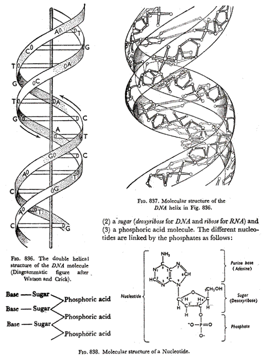
Although partly hypothetical, this concept is considered very reasonable. Even then many modifications have been suggested. For example, the DNA molecule probably very often contains several polynucleotide strands instead of the simple two assumed in the following clarification. Each nucleotide (Fig. 838) is composed of three different parts: (1) a nitrogen base.
In the DNA molecule only four nitrogenous bases (Fig. 835) are concerned—two Purines (Adenine and Guanine) and two Pyrimidines (Cytosine and Thymine). The four bases are symbolised as A, G, C and T respectively.
Although there are only four bases, the bases may be arranged in different orders in a polynucleotide chains (e.g., CCAGTTAC ………… or CAGTGA…………..) giving rise to hundreds of types of nucleic acid chains and thus causing different types of inherited characters. In RNA the bases are the same except that in place of Thymine there is Uracil, the bases being A, G, C & U. The RNA nucleotide is sometimes called ribotide and the chain polyribotide chain.
The big difference between RNA and DNA molecules is that while the RNA molecule is formed simply of single chains in DNA there are always two such parallel polynucleotide chains. Here, two such chains get linked—each adenine being linked with a thymine of the other chain through two H-atom bonds and similarly each cytosine getting linked to a guanine of the opposite chain by three H-atoms (Fig. 835).
Figs. 836, 837 and 839 also show this linking of two polynucleotide chains, the first diagrammatically and the latter two showing the actual molecular structure of a bit of the united chains. The molecular structure shows that the two chains cannot be straight so that they twist round each other giving the DNA molecule a double helical structure shown diagrammatically in Fig. 837 and with the molecular structure in Figs. 838 and 839. Such a molecule is very large and has a molecular weight of 5 to 10 million.
A number of such DNA molecules get joined lengthwise. The actual method of joining is not clear but, it is possibly through the proteins (histone and protamine) which are of basic nature and calcium also may play a role here. Thus, the long nucleoprotein fibrils are formed. The electron microscope has actually shown the presence of ‘micro-fibrils’, 100 to 200A (i.e., 10 to 20µµ) wide and coiled like a corkscrew inside the chromosome (Ambrose 1956).
The splitting and doubling of this polynucleotide paired threads of the chromosomes may also be explained on the basis of the DNA molecular structure. 
Splitting of the composite thread means somehow the hydrogen bonds between the nitrogenous bases break so that the two threads go apart (Fig. 840B) and immediately each nucleotide in the split threads attracts its opposite nucleotide from the free nucleotides present in the vicinity and the linked double chain is reformed (Fig. 840C) and ultimately, the whole chromosome gets doubled (Fig. 840D). This is called replication. We may, conclude that each fibril attracts identical material to itself, chain by chain and gene by gene, so that the original two strands of the helix take the places of two templates moulding the two new complementary strands.
The fixation of a complementary strand from the original double helix may also be compared with the developing of a photographic print from a negative (Fig. 840). When sufficient raw material accumulates, a new mitosis starts. During the mitotic cycle, the chromosomes lose water and thereby acquire a fibrous coat of nucleic acid (stainable matrix) as it coils and assumes the met-aphasic condensed appearance. Then it gains water, uncoils and ceases to be separately visible (loses stain-ability), i.e., reverts to the resting phase.
Endomitosis:
The chromosome number in a species of plants or animals is expected to be fixed. But, since the beginning of the current century spectacular discoveries have been made where it has been found that in the same plant there are patches of cells which show a different chromosome number. If a bud develops from such a plant, the new branch (which may become a separate plant) would show a different chromosome number. This is more common in plants which, propagate vegetatively. This difference may be due to non-disjunction (some chromosome pairs do not actually divide), somatic reduction etc. but most interesting is the case of endomitosis.
In endomitosis, the chromosomes of a somatic cell divide but the daughter chromosomes do not separate into daughter cell and the nuclear membrane does not break so that the -ultimate cell contains double the chromsomes (in case of complete endomitosis) or a different number (in case of partial endomitosis). The somatic patch may be said to be formed of endopolyploid cells and when perfect may contain 2n, 3n, 4n etc., cells.
Operon Concept:
There are thousands of genes in a cell every one of which secretes enzymes which give rise to different proteins. But, all these genes never act together and so no chaotic situation arises. The reason of this coordination had been considered a problem. Apparently, there must be some switching mechanism in the chromosomes which induces or stops the action of the genes. A hypothesis to explain this phenomenon has been presented by F. Jacob and J. Monod of the Paris Pasteur Institute in 1961 for which they were awarded a Nobel Prize (Biochemistry) in 1965.
They have suggested the operon model which pictures the operon which includes the gene codon along with similar genes which regulate, operate, repress or induce the action of the codon. The best worked out of the operons is the lac operon (lactose forming) of Escherechia coli which is a system of the codon TAC along with all its complicated controlling genes.
The Genes:
The ultimate unit which controls heredity is now called the gene. The word gene had been used by earlier Botanists but a clear picture came out only after the work by Morgan for which he was awarded a Nobel Prize.
Morgan stated his Theory of the Gene as follows:
“The theory states that the characters of the individual are preferable to paired elements (genes) , in the germinal material that are held together in a definite number of linkage groups; it states that the members of each pair of genes separate when the germ cells mature in accordance with Mendel’s first law, and in consequence each germ cell comes to contain one set only; it states that the members belonging to different linkage groups assort independently in accordance with Mendel’s second law; it states that an orderly interchange—crossing-over—also takes place, at times, between the elements in corresponding linkage groups; and it states that the frequency of crossing-over furnishes evidence of the linear order of the elements in each linkage group and of the relative position of the elements with respect to each other.”
What is the gene? It can be located on the chromosome and assigned to some particular locus, it can mutate and it can be hit by radiation rays like any other particle. The conclusion is that it is a particle and these particles are arranged on the chromonema thread in a linear order. Naturally, the chromomeres on the chrompnema threads were first supposed to be the genes or the seats of the genes.
When homologous chromosomes pair exactly identical chromomeres come to lie side by side. But further studies have shown that while some genes may be located in the chromomeres there are others in the interchromomeral regions. Moreover, recent workers doubt the suitability of considering the gene as a single particle.
The structures of the chromosome as now understood clearly defines the gene as a small segment of the polynucleotide chain consisting of hundreds or thousands of nucleotides, thus the gene can no longer be considered as a single unit—as a matter of fact, every nucleotide bit of it may act as a subunit.
Besides these subunits with the gene, there may be more elements within the genetic material affecting heredity which cannot be expressed by the assumption of a simple gene particle as shown by a linkage map. Nevertheless, although it has not yet been possible to identify these genes clearly under the optical microscope, their behaviour has made it possible to locate them and to speculate as to their dimensions and weights.
Muller (1947) located four genes on one of the Drosophila salivary gland chromosomes (Fig. 841) and calculated their mean length to be 1250Å, i.e., 125µm. Pontecarvo (1952) considered 4500A (450 fin) to be the gene size in the ascomycete Aspergillus nidulens. These are sizes of large virus particles.
It has been found that in some bacteria the bare nucleic acid may cause hereditary changes. The role of the protein in the chromosome is at yet uncertain. The nucleic acid of chromosomes is DNA but it has already been stated that in certain viruses RNA (ribose-nucleic acid—another type having the same characteristic as DNA) plays the same role and acts like genes. RNA, which is present in plant cytoplasm and nucleoli, is known to be connected with protein synthesis.
It has now been established that the nucleic acid DNA constitution the gene.
In support of this the following facts may be pointed out:
(i) The DNA content of a cell varies directly with the number of sets of chromosomes (haploid, diploid, triploid etc.), i.e., with the chromosome volume.
(ii) Different genetic problems can be best explained on the basis of the DNA constitution of the gene.
(iii) In virus (e.g. bacteriophage) it is the DNA core which causes hereditary transmission and not the protein casing.
(iv) Bacterial transduction:
In certain cases when the DNA material of a bacteriophage, on entering a bacterium does not destroy the latter but remains as a host inside the bacterium as a host side by side with the bacterial chromosome. The host DNA is then called a pro-phage. This pro-phage being smaller than the bacterial chromosome, behave as a gene and divides simultaneously with the bacterial chromosome but does not lose its identity. In this way the pro-phage is transferred from bacterium to bacterium and the phenomenon is called transduction. This behaviour of the pro-phage proves that DNA is the gene substance. Transduction has been put to further use in genetic engineering.
(v) Bacterial transformation:
Pneumococci producing disease have capsulated cells. Certain strains of Pneumococci do not possess capsules as they cannot synthesis the polysaccharide needed for it. These cannot cause any disease. If a non-capsulated strain be hybridised with a capsulated one, the offspring is capsulated- capsulation being dominant.
It has been found that if an extract of dead capsulated pneumococci be added to the non-capsulated strain the latter become capsulated. This is called transformation. Other instances of transformation have also been discovered. It has been found that the dead extract is at least 98% DNA. Hence, DNA is responsible for this transformation.
Considerable work has been done lately on the structure of the gene. Mendel’s concept was that the factors (now known as genes) were the ultimate units of heredity. But, lately it has been shown that the genes are divisible into subunits, i.e., units of mutation, units of recombination, etc. The DNA molecule of the chromosome contains a large number of genes which, again in turn, are quite large -and may be divided into finer subunits as the nucleotides or neucleotide triplets. Subunits are referred in terms of the different functions of the genes. Detailed studies of them have been done in the bacterium Escherichia coli.
Mention must be made of a very interesting and abnormal type of chromosomes which throws light on the structure of chromosomes and on genes. These are the giant salivary gland chromosomes of Diptera flies. The larvae of these flies (Drosophila, Chironoma, Bibio, Sciara, Rhyncosciara, Camptomya, etc.) show abnormally large cells with large nuclei (about 25µ. in diamater) in their salivary glands.
Similar cells are also found in their rectal epithelia and Malphighian tubes but the salivary glands are easier to dissect and study. These nuclei contain large chromosome, 50 to 200 times the normal size—some of them as much as half a millimetre long and visible under a simple dissecting microscope. Such chromosomes were noted by Balbiani in Chironoma as early as 1881 but their study only in the ’30s of the present century.
It is found that each such ‘chromosome’ is actually formed by a complete synapsis of the two homologous chromosomes along their entire lengths in the somatic cells. After this abnormal somatic synapsis cell division does not progress any further, – the cells remain permanently in the pro-phasis stage but the chromosomes increase in length enormously.
Moreover, it is seen that every chromosome shows distinct bands which are heavily stainable but less so in intercalary zones throughout the entire length (Fig. 841). The bands always occur at distinct places in a particular chromosome and are of definite widths. If a bit of a chromosome somehow gets lost, the lost zone can be easily detected by examining the pattern of the zones. Thus each chromosome has a definite and constant pattern which may be called a. biological spectrum. On the large chromosome of Drosophila there are more than 2000 distinct bands along its length.
In Drosophila, the four chromosomes remain adhering to each other at the centromeres. All the heterochromatic material from near the centromeres of all the chromosomes (as also the whole Y-chromosome of the male fly, which is formed almost entirely of heterochromatin) fuse together to form a chromo-centre. From this chromo-centre the four chromosomes spread out like the arms of an octopus (Fig. 842)—the two long chromosomes II and III, having centromeres in the middle, show two arms each. In other Diptera flies the chromosomes remain separate from one another in the nucleus.
Naturally, these bands have been studied closely. Each band seems to be formed of one or more Lumps of chromatic granules extending across the cylindrical double-chromosomes. These are the regions where nucleic acid accumulates greatly and they may be the seats of the genes.
If a fly becomes mutant by the loss or deletion of a bit of a chromosome and if a hybrid be obtained between this mutant and a normal fly, the salivary gland chromosome where the two homologous chromosomes are united show the ‘deletion’ in one half of it and this enables the location of the genes.
It has been supposed that the bands on the salivary gland chromosomes represent the chromomeres of the normal chromonema. Somehow or other the original chromonema goes on dividing longitudinally repeatedly so that the salivary gland chromosome shows not two or four chromonemata as in a normal chromosome but a large number of them. This is termed polyteny. Some polytene chromosomes have been estimated to contain more than a thousand chromonemata. Such repeated division of chromonema threads without actual cell division is termed endomitosis.
As all these chromonema threads are identical, they have the chromomeres at identical spots which press on one another as the chromonemata are closely pressed together. So many chromomeres at one place appear as a band or disc on the chromosome which is completely uncoiled. Some authorities, however, think that such a structure is also possible by the swelling of the four normal chromonemata instead of undergoing actual polyteny.
In Genetics, genes are symbolised in the same way as unit characters of Mendel by letters, e.g., T and d. It is customary to use capital letters for dominant genes and small letters for recessive genes. Nowadays, the same letter or letters are used for pairs of allelomorphic genes, e.g., A and a form one pair and Pr with pr form another. Dominant genes found in the wild type are frequently do noted by the common symbol +.


