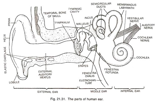
In this article we will discuss about the structure and functions of human ear.
Structure of Ear:
Each ear consists of three portions:
(i) External ear,
(ii) Middle ear and
(iii) Internal ear.
1. External Ear:
It comprises a pinna, external auditory meatus (canal) & tympanic membrane.
(i) Pinna:
The pinna is a projecting elastic cartilage covered with skin. Its most prominent outer ridge is called the helix. The lobule is the soft pliable part at its lower end composed of fibrous and adipose tissue richly supplied with blood capillaries. It is sensitive as well as effective in collecting sound waves.
(ii) External Auditory Meatus:
It is a tubular passage supported by cartilage in its exterior part and by bone in its inner part. The meatus (canal) is internally lined by hairy skin (stratified epithelium) and ceruminous glands (wax glands). The latter are modified sweat glands which secrete a waxy substance— the cerumen (ear wax) which prevents the foreign bodies entering the ear.
(iii) The tympanic membrane (tympanum):
Separates the tympanic cavity from the external auditory meatus. It is thin and semi-transparent, almost oval, though somewhat broader above than below. The central part of the tympanic membrane is called the umbo. The handle of the malleus is firmly attached to the membrane’s internal surface.
Functions of External Ear:
It directs sound waves towards the tympanic membrane. The sound waves produce pressure changes over the surface of the tympanic membrane. The cerumen (ear wax) prevents the entry of the foreign bodies into the ear.
2. Middle Ear:
It includes the following:
(i) The tympanic cavity, filled with air is connected with the nasopharynx through the Eustachian tube (auditory tube), which serves to equalize the air pressure in the tympanic cavity with that on the outside.
(ii) There is a small flexible chain of three small bones called ear ossicles— the malleus (hammer shaped), the incus (anvil shaped) and the stapes (stirrup shaped). The malleus is attached to the tympanic membrane on one side and to the incus on the other side.
The incus in turn is connected with the stapes, which is attached to the oval membrane covering the fenestra ovalis (oval window) of the inner ear. Malleus is the largest ossicle, however, stapes is smallest ossicle. Stapes is also the smallest bone in the body.
(iii) Two skeletal muscles, the tensor tympani attached to the malleus and the stapedius attached to the stapes, are also present in the middle ear. Stapedius is the smallest muscle in the body.
(iv) The middle ear is connected with the inner ear through two small openings closed by the membranes. These openings are (a) fenestra ovalis (oval window) as mentioned above and (b) fenestra rotunda (round window).
The fenestra ovalis is covered by foot plate of the stapes. The fenestra rotunda is enclosed by a flexible secondary tympanic membrane. The latter is responsible for equalizing the pressure on either side of the tympanic membrane.
Functions of Middle ear:
(i) Due to the pressure changes produced by sound waves, the tympanic membrane vibrates, i.e., it moves in and out of the middle ear. Thus the tympanic membrane acts as a resonator that reproduces the vibration of sound,
(ii) It transmits sound waves from external to the internal ear through the chain of ear ossicles,
(iii) The intensity of sound waves is increased about twenty times by the ear ossicles. It may be noted that the frequency of sound does not change and
(iv) From the tympanic cavity extra sound is carried to the pharynx through Eustachian tube.
3. Internal Ear:
There is a body cavity on each side enclosed in the hard periotic bone which contains the perilymph. The later corresponds to the cerebrospinal fluid. A structure, the membranous labyrinth floats in the perilymph. The membranous labyrinth consists of three semicircular ducts, utricle, saccule, endolymphaticus and cochlea.
(i) Semicircular Ducts:
There are present three semicircular ducts; the anterior, the posterior and the lateral semicircular ducts. They arise from the utricle. The anterior and posterior semicircular ducts arise from crus commune.
Each semicircular duct is enlarged at one end to give rise to a small rounded ampulla. The anterior and lateral semicircular ducts bear ampullae at their anterior ends, while the posterior duct contains an ampulla at its posterior end.
Each ampulla contains a sensory patch of cells, the crista Each crista consists of two kinds of cells, the sensory and supporting cells. The sensory cells bear long sensory hairs at their free ends and nerve fibres at the other end. The sensory hairs are partly embedded in a gelatinous mass, the cupula. The cristae are concerned with balance of the body.
(ii) Utricle, Endolymphaticus and Saccule:
The utricle is a dorsally placed structure to which all the three semicircular ducts are connected. The saccule is a ventrally situated structure which is joined with the utricle by a narrow utriculosaccular duct. From this duct a long tube, the ductus endolymphaticus arises which ends blindly as the saccus
endolymphaticus. Both utricle and saccule contain sensory patches, the maculae. A macula comprises sensory and supporting cells similar to those of the crista. The hair are not actually motile and are embedded in a gelatinous membrane, the otolith membrane in which there are also found very small crystals of calcium carbonate, the otolith. The cristae and maculae are the receptors of balance.
Both cristae and maculae are concerned with balance.
(iii) Cochlea:
It is the main hearing organ which is connected with saccule by a short ductus reuniens leading from the saccule. It is spirally coiled that resembles a snail shell in appearance. It tapers from a broad base to an almost pointed apex.
Internally it consists of three fluid filled chambers or canals, the upper scala vestibuli, lower scala tympani, and the middle scala media (cochlear duct). Both scala vestibuli and scala tympani are filled with perilymph. However scala media is filled with endolymph. Both the scala vestibuli and scala tympani are connected with each other at the apex of the cochlea by a small canal, the helicotrema.
It is important to mention that near the base of the scala vestibuli the wall of the membranous labyrinth comes in contact with the fenestra ovalis, while at the lower end of the scala tympani lies the fenestra rotunda.
The scala media is the most important canal or channel of the cochlea. It bears an upper membrane, the Reissner’s membrane, and lower membrane, basilar membrane. On the basilar membrane a sensory ridge, the organ of Corti is present.
The organ of Corti consists of outer hair cells, inner hair cells, inner pillar cells, outer pillar cells, tunnel of Corti, phalangeal cells (cells of Deiters), cells of Hensen and cells of Claudius.
The sensory hairs project from the outer ends of the hair cells into the scala media, while from the inner end of the cells nerve fibres arise, which unite to form the cochlear nerve. The tectorial membrane overhangs the sensory hair in the scala media. Its properties are to determine the patterns of vibration of sound waves.
Functions of Ear:
The ear performs the functions of hearing and balancing (equilibrium).
1. Mechanism of Hearing:
The sound waves are collected by the external ear up to some extent. They pass through the external auditory meatus to the tympanic membrane which is caused to vibrate. The vibrations are transmitted across the middle ear by the malleus, incus and to the stapes bones. The latter fits into the fenestra ovalis. The perilymph of the internal ear receives the vibrations through the membrane covering, the fenestra ovalis.
From the perilymph the vibrations are transferred to the scala vestibuli of cochlea and then to scala media through Reissner’s membrane. Thereafter, the movements of endolymph and tectorial membrane stimulate the sensory hairs of the organ of Corti.
The impulses thus received by the hair cells are carried to the brain (temporal lobe of each cerebral hemisphere) through the auditory nerve where the sensation of hearing is felt (recognised).
It is evident that the external and middle ears serve to transmit sound waves to the internal ear. It is in the internal ear that the transformation of the vibrations into nerve impulses for relay to the brain takes place. During loud sound, some sound waves are transferred from scala vestibuli to scala tympani through helicotrema.
From scala tympani the sound waves are transmitted to the tympanic or middle ear cavity through the membrane covering the fenestra rotunda. From the tympanic cavity the sound waves are transferred to the pharynx through the Eustachian tube.
2. Equilibrium:
The semicircular canals, utricle and saccule of membranous labyrinth are the structures of equilibrium (balancing). Whenever the animal gets tilted or displaced the hair cells of the cristae and maculae are stimulated by the movement of the endolymph and otolith.
The stimulus is carried to the brain through the auditory nerve and the change of the position is detected by the medulla oblongata of the brain. After that, the brain sends impulses (messages) to the muscles to regain the normal conditions.
i. Meniere’s Disease:
Spinning or whirling vertigo (dizziness) is characteristic of meniere’s disease.
ii. Ottis Media:
This is an acute infection of the middle ear caused mainly by bacteria and associated with infection of the nose and throat.


