
In this article we will discuss about the mechanisms of blood circulation in humans:- 1. Coronary Circulation 2. Cerebral Circulation 3. Microcirculation 4. Pulmonary Circulation 5. Fetal Circulation 6. Splanchnic Circulation 7. Intestinal Circulation.
1. Coronary Circulation:
Coronary circulation is the circulation of blood in the blood vessels of the heart muscle (myocardium). Normal coronary blood flow at rest is about 60-80 ml/100 gm/min (or) 250 ml/min blood supply.
In general there are two main coronary arteries, the left and right:
i. Right coronary artery
ii. Left coronary artery.
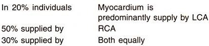
Both of these arteries originate from the beginning (root) of the aorta, immediately above the aortic valve. The left coronary artery originates from the left aortic sinus, while the right coronary artery originates from the right aortic sinus.
The right coronary artery branches to smaller arteries including the marginal, which leads down the margin or edge of the right ventricle. The main portion of the right coronary artery proceeds to the back of the heart becoming the posterior interventricular.
The left coronary artery divides to form the circumflex which curves to the back of the heart, and the anterior interventricular which descends between the two ventricles. The arteries anastomose to provide collateral circulation to the ventricular myocardium.
Coronary veins drain the myocardium from the anterior interventricular area through the great cardiac (or coronary) vein, from the right atrial area through the small cardiac vein, and from the posterior interventricular area through the middle cardiac vein. All of these come together to form the coronary sinus which drains directly into the right atrium, the only systemic venous drainage not through the vena cavae. There are also anterior cardiac veins and thebesian veins drain directly into the cardiac chambers.
The left and right coronary arteries and their branches lie on the surface of the heart, and therefore, are sometimes referred to as the epicardial coronary vessels. These vessels distribute blood flow to different regions of the heart muscle. The arterioles branch into numerous capillaries that lie adjacent to the cardiac myocytes. A high capillary-to-cardiomyocyte ratio and short diffusion distances ensure adequate oxygen delivery to the myocytes and removal of metabolic waste products from the cells (e.g. CO2 and H+).
2. Cerebral Circulation:
The brain, though representing 2% of the total body weight, it receives one-fifth of the resting cardiac output. The amount of blood that the cerebral circulation carries is known as cerebral blood flow.
Blood Supply:
This blood supply is carried by the two internal carotid arteries (ICA) and the two vertebral arteries that anastomose at the base of the brain to form the circle of Willis.
Carotid arteries and their branches (referred to as the anterior circulation) supply the anterior portion of the brain while the vertebrobasilar system (referred to as posterior circulation) supplies the posterior portion of the brain.
Venous Drainage:
Venous blood flows peripherally via superficial cerebral veins and centrally via the deep cerebral veins into the venous sinuses which drain into the internal jugular vein. The cerebral veins are thin-walled and have no valves. There are numerous venous connections between cerebral veins and dural sinuses and venous systems of the meninges, skull, scalp and nasal sinuses so facilitating propagation of thrombus or spread of infection between these vessels.
3. Microcirculation:
Structure:
The microcirculation is a term used to describe the small vessels in the vasculature which are embedded within organs and are responsible for the distribution of blood within tissues.
The vessels on the arterial side of the microcirculation are called the arterioles. Arterioles carry the blood to the capillaries. Blood flows out of the capillaries into the venules. The blood flows from venules into the veins. In addition to these blood vessels, the microcirculation also includes lymphatic capillaries and collecting ducts.
Arterioles:
i. Small pre-capillary resistance vessels (10-50 µ) composed of an endothelium surrounded by one or more layers of smooth muscle cells.
ii. The endothelium provides a smooth surface for the flow of blood and regulates the movement of water and dissolved materials in the plasma between the blood and the tissues.
iii. The endothelium also produces molecules that discourage the blood from clotting unless there is a leak.
iv. The smooth muscle cells can contract and decrease the size of the arterioles and thereby regulate blood flow and blood pressure.
v. Richly innervated by sympathetic adrenergic fibers and highly responsive to sympathetic vasoconstriction via both α1 and α2 post-junctional receptors.
vi. Represent a major site for regulating systemic vascular resistance.
vii. Primary function within an organ is flow regulation, thereby determining oxygen delivery and the washout of metabolic byproducts.
viii. Regulate, in part, capillary hydrostatic pressure and therefore influence capillary fluid exchange.
Capillaries:
i. Small exchange vessels (6-10 µ) composed of highly attenuated (very thin) endothelial cells surrounded by basement membrane. There is no smooth muscle.
ii. This layer is so thin that molecules such as oxygen, water and lipids can pass through them by diffusion and enter the tissues. Waste products such as carbon dioxide and urea can diffuse back into the blood to be carried away for removal from the body.
iii. They are very prevalent in the body; total surface area is about 6,300 square meters. Because of this, no cell is very far from a capillary, no more than 50 micrometers away.
iv. The “capillary bed” is the network of capillaries present throughout the body. These beds are able to be “opened” and “closed” at any given time, according to need. This process is called auto-regulation and capillary beds usually carry no more than 25% of the amount of blood it could hold at any time. The more metabolically active the cells, the more capillaries it will require to supply nutrients.
Three structural classifications are given below:
a. Continuous (Non-Fenestrated):
i. Found in muscle, skin, lung, central nervous system
ii. Basement membrane is continuous and intercellular clefts are tight (i.e. have tight junctions); these capillaries have the lowest permeability.
b. Fenestrated:
i. Found in exocrine glands, renal glomeruli, and intestinal mucosa.
ii. Perforations (fenestrae) in endothelium result in relatively high permeability.
c. Sinusoidal (Discontinuous):
i. Found in liver, spleen, bone marrow
ii. Large intercellular gaps and gaps in basement membrane result in extremely high permeability.
Venules:
i. Small vessels (10-50 µ) composed of endothelial cells surrounded by basement membrane (smallest post-capillary venules) and smooth muscle (larger venules).
iii. Fluid and macromolecular exchange occur most prominently at venular junctions.
iv. Sympathetic innervation of larger venules can alter venular tone which plays role in regulating capillary hydrostatic pressure.
Terminal Lymphatics:
i. Composed of endothelium with intercellular gaps surrounded by highly permeable basement membrane and are similar in size to venules ― terminal lymphatics end as blind sacs.
ii. Larger lymphatics also have smooth muscle cells.
iii. Spontaneous and stretch-activated vasomotion is present which serves to “pump” lymph.
iv. Sympathetic nerves can modulate vasomotion and cause contraction.
v. One-way valves direct lymph away from the tissue and eventually back into the systemic circulation via the thoracic duct and subclavian veins (2-4 liters/day returned).
Functions:
The main functions of the microcirculation include the:
i. Regulation of blood flow and tissue perfusion. Flow is determined by the diameter and the length of the vessels of the microcirculation. The Hagen-Poiseuille equation predicts the flow of blood through the vessels.
ii. Regulation of blood pressure, by capillary fluid shift mechanism.
iii. Regulation of tissue fluid (swelling or edema), by capillary exchange of water. The starling equation is an equation that describes the roles of hydrostatic and oncotic forces (the so-called Starling forces) in the movement of fluid across capillary endothelium.
iv. Delivery of oxygen and other nutrients and removal of CO2 and other metabolic waste products, by capillary exchange of solutes. Small solutes move across the endothelium by passing through the spaces formed by the tight junctions formed where the edges of adjacent endothelial cells abut.
v. Regulation of body temperature. Triple response (of Lewis) ― It is a physiological reaction of the skin to stroking with a blunt instrument ― first a red line develops at the site of stroking, due to capillary dilatation because of release of histamine or a histamine-like substance, then a flare develops, i.e. redness in the surrounding area due to arteriolar dilatation mediated by axon reflex, and lastly a wheal is formed as a result of exudation of fluid from capillaries and venules.
4. Pulmonary Circulation:
The quantity of blood flowing through the lungs is essentially equal to that flowing through the systemic circulation. The pulmonary trunk extends only 5 cm beyond the apex of the right ventricle and then divides into the right and left main branches, which supply blood to the two respective lungs. The pulmonary artery is also thin with a wall thickness one-third that of the aorta. The pulmonary arterial branches are all very short.
However, all the pulmonary arteries, smaller arteries and arterioles have larger diameter than their counterpart systemic arteries. Pulmonary arterioles subdivide to form network of pulmonary capillaries which surrounds the alveoli and are sandwiched between their walls. Alveoli are kept in a basket of capillaries.
Effective wall surface area at rest is 60 m2 and transit time across the capillaries is 0.8 sec. During heavy exercise, it increases to 90 m2. Pulmonary venules and veins receive oxygenated blood from the capillaries; they join to form 4 main veins which finally open into the left atrium.
The pulmonary veins like the pulmonary arteries are short but their distensibility characteristics are similar to those of the veins in the systemic circulation.
Bronchial Vessels:
Blood also flows to the lungs through several bronchial arteries, amounting to about 1-2% of cardiac output. This bronchial arterial blood is oxygenated blood. It supplies the supporting tissues of the lungs, including the connective tissue, septa and large and small bronchi. From this, deoxygenated blood enters into the pulmonary veins and enters the left atrium, rather than passing back to the right atrium.
Therefore, the flow into the left atrium and left ventricular output are about 1-2% greater than the right ventricular output.
Lymphatics:
Lymphatics extend from all the supportive tissue of the lung, beginning in the connective tissue spaces that surrounded the terminal bronchioles and coursing to the hilum of the lung and hence mainly into the right lymphatic duct. Particulate matter entering the alveoli is partly removed by way of these channels, and plasma protein leaking from the lung capillaries is also removed from the lung tissue, thereby helping to prevent edema.
5. Fetal Circulation:
The circulation of the fetus shows a number of differences from that of the postnatal infant. The fetal lungs are functionally inactive, and the fetus depends completely on the placenta for O2 and nutrient supply.
Specific Anatomical Structure of the Fetal Circulation:
Oxygenated blood from the placenta passes through the umbilical vein to the liver. A minor fraction passes through the liver and a major fraction bypasses the liver to the inferior vena cava through the ductus venous. In the inferior vena cava, blood from the ductus venosus joins blood returning from the lower trunk and extremities; this combined stream is in turn joined by blood from the liver through the hepatic veins. The streams of blood maintain their identity in the inferior vena cava and are divided into 2 streams of unequal size by the edge of the interatrial septum.
The larger stream, which is mainly blood from the umbilical vein, is shunted to LA through foramen ovale.
The other stream passes into right atrium, where it is joined by superior vena caval blood returning from the upper parts of the body and by blood from myocardium.
In contrast to the adult, in whom the right and left ventricles pump in series; in fetus the ventricles operate in parallel.
Because of the large pulmonary resistance, less than 1/3rd of right ventricle output goes through the lungs. The remainder passes through the ductus arteriosus from the pulmonary artery to the aorta at a point distal to the origin of the arteries to the head and upper extremities. Flow from pulmonary artery to aorta occurs because pulmonary arterial pressure is 5 mm Hg higher than that of aorta.
The large volume of blood coming through foramen ovale into left atrium is joined by blood returning from the lungs and is pumped out by left ventricle into the aorta.
About 1/3rd of aortic blood goes to the head, upper thorax and arms; remaining 2/3rds go to the rest of the body and the placenta (via 2 umbilical arteries).
i. Fetal blood leaving the placenta is 80% saturated, but the saturation of the blood passing through the foramen ovale is reduced to 67% by mixing with de-saturated blood returning from the lower part of the body and the liver. Addition of the de-saturated blood from the lungs reduces the O2 saturation of left ventricular blood to 62%, which is the level of saturation of the blood reaching the head and upper extremities.
The blood in the right ventricle, a mixture of de-saturated superior vena caval blood, coronary venous blood, and inferior vena caval blood, is only 52% saturated with O2. when the major portion of the blood traverses the ductus arteriosus and joins that pumped by the left ventricle, the resultant O2 saturation of blood traveling to the lower part of the body and back to the placenta is 58% saturated.
Thus, it is apparent that the tissue receiving blood of the highest O2 saturation are the liver, heart and upper parts of the body, including the head.
At the placenta, the chorionic villi dip into the maternal sinuses, and O2, CO2, nutrients, and metabolic waste products exchange across the membranes. The barrier to exchange is quite large, and the equilibrium of O2 tension between the two circulations is not reached at normal rate of blood flow. Therefore, the O2 tension of the fetal blood leaving the placenta is very low.
Since, the fetal hemoglobin has higher O2 affinity, oxygen dissociation curve is shifted to the left.
Features:
1. All the blood is eventually transferred from the right heart to the aorta, as in the adult. But unlike the adult, it takes three routes to do so. The adult route (via lungs) is taken by only about 13% of right atrial blood. Other 2 unique routes are foramen ovale and ductus arteriosus. They close soon after birth.
2. The aortic flow is distributed to the whole body as in the adult. But a unique artery arising from fetal aorta is umbilical artery which carries blood to placenta. After improving to O2 content in the placenta, the blood returns to the fetus through the umbilical vein. Thus, the placenta acts like the lungs of the adult. It also acts like the gut and kidneys in the sense that it adds nutrients to and also removes waste products from the fetal blood.
6. Splanchnic Circulation:
The splanchnic circulation is composed of gastric, small intestinal, colonic, pancreatic, hepatic, and splenic circulations, arranged in parallel with one another. It receives about 1500 ml of blood at rest (30% of cardiac output).
The three major arteries that supply the splanchnic organs are celiac, superior and inferior mesenteric, which give rise to smaller arteries that anastomose extensively:
i. Celiac artery supplies 200 ml to stomach and spleen and 500 ml to liver via hepatic artery.
ii. Superior mesenteric artery supplies 500 ml to pancreas, small intestine and parts of colon.
iii. Inferior mesenteric artery supplies 300 ml to colon.
The Hepatic Vascular System:
The circulatory system of the liver is unlike that seen in any other organ. Of great importance is the fact that a majority of the liver’s blood supply is venous blood!
The pattern of blood flow in the liver can be summarized as follows:
1. Roughly 75% of the blood entering the liver is venous blood from the portal vein. Importantly, all of the venous blood returning from the small intestine, stomach, pancreas and spleen converges into the portal vein. One consequence of this is that the liver gets “first pickings” of everything absorbed in the small intestine, which, as we will see, is where virtually all nutrients are absorbed.
2. The remaining 25% of the blood supply to the liver is arterial blood from the hepatic artery.
Terminal branches of the hepatic portal vein and hepatic artery empty together and mix as they enter sinusoids in the liver. Sinusoids are distensible vascular channels lined with highly fenestrated or “holey” endothelial cells and bounded circumferentially by hepatocytes. As blood flows through the sinusoids, a considerable amount of plasma is filtered into the space between endothelium and hepatocytes (the “space of Disse”), providing a major fraction of the body’s lymph.
Blood flows through the sinusoids and empties into the central vein of each lobule. Central veins coalesce into hepatic veins, which leave the liver and empty into the vena cava.
Hepatic Blood Volume and Reservoir Function:
The liver receives approximately 30% of resting cardiac output and, is therefore, a very vascular organ. The hepatic vascular system is dynamic, meaning that it has considerable ability to both store and release blood—it functions as a reservoir within the general circulation.
In the normal situation, 10-15% of the total blood volume is in the liver, with roughly 60% of that in the sinusoids. When blood is lost, the liver dynamically adjusts its blood volume and can eject enough blood to compensate for a moderate amount of hemorrhage. Conversely, when vascular volume is acutely increased, as when fluids are rapidly infused, the hepatic blood volume expands, providing a buffer against acute increases in systemic blood volume.
7. Intestinal Circulation:
In the intestines the arteries enter the wall and circle round the gut and send branches to the muscular layers, mucosa and the villi. The mucosa receives more blood than other parts. In the villi, the artery and vein are close to and parallel to each other with blood flow in opposite direction (countercurrent). Blood flow responds to changes in metabolic activity.
Skeletal Muscle Blood Flow:
Skeletal muscle accounts for about 20% of cardiac output and systemic vascular resistance. During extreme physical exertion, more than 80% of cardiac output can be directed to contracting muscles; therefore, skeletal muscle resistance becomes the primary determinant of systemic vascular resistance during exercise.
Blood Flow:
At rest, skeletal muscle blood flow may be 1-4 ml/min per 100 g. Maximal blood flow may reach 50-100 ml/min per 100 g depending upon the muscle type. Therefore, blood flow can increase 20 to 50- fold with maximal vasodilation or active hyperemia.
Coordinated, rhythmical contractions (e.g. running) enhance blood flow by means of the skeletal muscle pump mechanism.
Regulation:
1. Sympathetic innervation produces vasoconstriction through α1 and α2-adrenoceptors located on the vascular smooth muscle. There is a significant amount of sympathetic tone at rest so that abrupt removal of sympathetic influences (e.g. by using an alpha-adrenoceptor blocker) can increase resting flow 2 to 3-fold.
Vascular β2-adrenoceptors produce vasodilation when stimulated by agonists such as epinephrine. There is evidence for sympathetic cholinergic innervation of skeletal muscle arteries, particularly large arteries. Activation of these autonomic nerves during exercise can cause neural-mediated vasodilation through the release of acetylcholine binding to muscarinic receptors.
2. There is a close coupling between oxygen consumption and blood flow.
3. Blood flow is strongly determined by local regulatory (tissue and endothelial) factors such as tissue hypoxia, adenosine, K+, CO2, H+, and nitric oxide. During exercise, these local regulatory mechanisms override the sympathetic vasoconstrictor influences (termed functional sympatholysis).
4. Skeletal muscle blood flow shows a moderate degree of auto-regulation.
5. Like the coronary circulation, muscle blood flow can be significantly compromised by extravascular compression that occurs during strong muscular contractions, especially during sustained tetanic contractions.