The following points highlight the eleven main organelles that eukaryotic cytoplasm possess. The organelles are: 1. Endoplasmic Reticulum (ER) 2. Golgi Complex 3. Lysosomes 4. Peroxisomes 5. Glyoxysomes 6. Spherosomes 7. Vacuole 8. Centrosome 9. Ribosomes 10. Chloroplast 11. Mitochondria.
Organelle # 1. Endoplasmic Reticulum (ER):
Endoplasmic reticulum is a highly convoluted sheet of membranes extending from the nuclear membrane. About 30-60% of the total membranes of the cell are those forming ER. The interior space within ER membrane folds is called the “lumen” (Fig. 2.2, 2.11).
ER is divided into two types:
(1) Rough ER and
(2) Smooth ER (Fig. 2.2).
Both the forms are the part of the same membrane sheet and are inter-convertible. In rough ER, ribosomes are attached to the cytoplasmic surface of the ER membranes which gives them the typical ‘rough’ appearance. The smooth ER, on the other hand, is devoid of ribosomes. Rough ER is predominant in metabolically active cells, particularly those involved in protein synthesis.
Extensive smooth ER is present in the cells which are involved in metabolism of steroids, drugs and toxic substances. The cytoplasm is divided by ER into cell sap (cytosol) and cisternal space (lumen). The lumen is continuous with internal cavities of Golgi complex, lysosomes and perinuclear space etc. The cytosol contains soluble enzymes, tRNAs and free ribosomes.
The function of ER may be summarized as follows:
(i) Rough ER provides the place for ribosome attachment which are the sites of protein synthesis.
(ii) Smooth ER is the place where most of the lipids and polysaccharides are synthesized.
(iii) ER provides a channel for the transport of proteins and other materials to different parts of the cell.
(iv) The ER also provides channels for secretion of proteins and other products out side the cell, following the route of ER à Golgi membranes à vesicles à Plasma membrane (Fig. 2.11).
Organelle # 2. Golgi Complex:
Golgi complex is called dictyosome in plant cells. A Golgi body consists of a stack of 20 or more flattened smooth sacs enclosed by membrane; the sacs are curved to assume a convex appearance, called the cts-face, on one side and a concave, known as the trans-face, on the other (Fig. 2.11).
There are several vesicles and membranous channels associated with the Golgi complex. It originates from the ER. The enzyme “thiamine pyrophosphatase” is present in the Golgi complex; it serves as a marker because it is rarely found in any other organelle. Nucleoside phosphatase is also used as a marker for the complex.
Function of Golgi Complex:
Golgi complex produces lysosomes and secretory granules. Newly synthesized proteins, lipids, carbohydrates in association with the ER, are transported to the bioregion of the complex. They are sorted out and suitably modified in this complex; they are then packed in vesicles at trans region.
Some of the granules contain hydrolytic enzymes and form lysosomes, while others are secreted; the secretion can be described in the following steps:
(i) Transport of Substances from ER:
Proteins and other products are enclosed in uncoated vesicles called transitional vesicles. These vesicles are formed in the ER; they migrate to the Golgi complex and fuse with the c/s-face of the complex. This step requires energy which is provided by ATP.
(ii) Selection and Modification of the Products:
Secretory proteins move towards the trans- face. The substances involved in lysosomes and plasma membrane also pass through the same route in Golgi complex. Modification of proteins occur by addition of fatty acid residues (to form lipoproteins) or polysaccharides (to form glycoproteins).
(iii) Packing of Secretary Products into Granules:
The secretory products are concentrated in membrane bound vesicles (granules) of different sizes produced on the trans-face. Fusion of small vesicles occurs to produce larger vesicles called Zymogen granules.
(iv) Release of the Secretory Products:
The discharge of secretory substances may be mediated by Ca2+ ions. The concentration of Ca2+ ions increases during this process. Secretory granules reach the plasma membrane and fuse with it thereby secreting the products contained in the vesicles (exocytosis). This process should result in an expansion of the plasma membrane.
However, there is neither an expansion of the plasma membrane nor a reduction in the Golgi membranes. Probably, the plasma membrane buds off vesicles which move to the Golgi complex; this would prevent the expansion of the plasma membrane due to the continuous fusion of granules, particularly in the secretory cells. (Fig. 2.11).
Organelle # 3. Lysosomes:
The term lysosome (= hydrolytic particle) was coined by de Duve in 1955. Lysosomes are 100- 200 nm vesicles enclosed by a membrane.
They are classified into two types:
(i) Primary lysosomes and
(ii) Secondary lysosomes.
Primary lysosomes contain hydrolytic enzymes; they are yet to participate in the digestive activity. When a primary lysosome fuses with an endosome (a vesicle containing food particle etc.), it gives rise to a secondary lysosome or hetero-phagic lysosome. Lysosomes contain the following hydrolytic enzymes.
a. Nucleases (acid ribonuclease, acid de-oxy-ribonuclease).
b. Phosphatases (acid phosphatase, acid pyrophosphatase, phosphoprotein phosphatase, phosphodiesterase).
c. Lipases (esterase, triglyceride lipase, phospholipase, galactocerebrosidase, sphingomyelinase).
d. Glycosidases (P-galactosidase, P-xylosidase, p-glucosidase, a-glucosidase, a- mannosidase, a-N-acetyl hexosaminidase, lysozyme, hyaluronidase).
e. Proteases and peptidases (peptidase, collagenase arylamidase).
f. Sulfatases (arylsulfatase).
However, all the lysosomes do not contain all the enzymes.
Production of Lysosomes:
The hydrolytic enzymes synthesized in association with ER bound ribosomes enter the lumen of the ER. Then oligosaccharide chains are transferred to the polypeptide chains. The phosphorylation of mannose (in the oligosaccharide chain) occurs to form mannose-6-phosphate, and the ‘enzyme-mannose-6-phosphate’ complex migrates to the Golgi complex.
The lysosomal enzymes are identified by the receptors in Golgi complex due to presence of mannose-6-phosphate. These enzymes are then packed into vesicles which become lysosomes.
Organelle # 4. Peroxisomes:
Peroxisomes are the vesicles containing enzymes that are involved in respiration (other than mitochondria). The most common enzyme present in peroxisomes is catalase. Peroxisomes occur primarily in leaves and seeds of plants, in lower eukaryotic organisms, such as, yeast, algae and fungi, and in the liver and kidney cells of vertebrates.
They contain a number of oxidizing enzymes like catalase, D-amino acid oxidase, glycolate oxidase, urate oxidase, malate dehydrogenase, fatty acid β-oxidation, L-α-hydroxy acid oxidase, citrate synthase, malate, synthase, isocitratelyase and glyoxylatereductases etc.
Peroxisomes from different tissue sources vary in the enzymes present. These enzymes break down the concerned organic compounds and produce H2O2 (taxic substance) utilizing molecular oxygen. The catalase breaks down H2O2 as follows.
In the above reaction, one molecule of oxygen and two molecules of water arc produced.
The main functions of peroxisomes are:
(i) Toxic substances, such as, ethanol, methanol and farm-aldehyde etc.. are broken down by the enzymes, and hydrogen peroxide (H2O2) is produced.
(ii) Peroxisomes utilize O2 in respiration and thus they protect the cell from adverse effects of high concentrations of O2.
(iii) During photosynthesis, excess production of O2 is utilized by peroxisomes in a process called photorespiration.
(iv) Peroxisomes are also involved in the reproduction of cytoplasmic NAD+.
However, the energy produced by oxidation in peroxisomes is lost since it is not stored.
Biogenesis of peroxisomes occurs by expansion and budding of the existing peroxisomes. The peroxisomal enzymes are produced on the free ribosomes present in cytosol; these enzymes are then included in the peroxisomes.
Organelle # 5. Glyoxysomes:
They are special types of vesicles which contain enzymes for glyoxalate cycle. Glyoxysomes are found in plants only. They also contain catalase and some other oxidases. In glyoxalate cycle, carbohydrates are synthesized from the products of fatty acid oxidation. It differs from the Krebs’s cycle in respect of the steps that bypass the CO2-evolving steps.
Organelle # 6. Spherosomes:
These are membrane-bound vesicles with a diameter of 0.5 to 1 pm which show acid hydrolase activity. They are highly refractive particles and are confined to plants. The main function of spherosomes is in storing and mobilizing reserve lipids.
Organelle # 7. Vacuole:
Vacuoles of different sizes are found in plant cells. They are enclosed by a membrane called tonoplast (Fig. 2.2). Immature cells contain small vaculoes scattered in the cytoplasm but in mature cells they fuse to form a large vacuole which may occupy 80-90% of the cytoplasmic volume.
Vacuoles contain sugars, salts, acids, alkaloids, anthocyanin pigments, glycosides, droplets of fats and oils and other substances such as tannin etc. In mature cells, calcium oxalate crystals are also found in the vacuoles. Vacuoles have the following two main functions.
(i) It functions as a water reservoir for the cell and maintains the cellular structure by exerting pressure.
(ii) It acts as a place for the storage of materials and cellular wastes. Through the tonoplast, the hydrolytic enzymes are secreted into the vacuole. ER and Golgi complexes fuse with tonoplast and release their materials in the vacuole. Hydrolytic enzymes degrade the waste material into simpler substances which may be absorbed by the cell.
Organelle # 8. Centrosome:
Centrosome is located adjacent to the nucleus in animal cells, but not in plants. Each centrosome contains two cylindrical centrioles of 150-200 nm diameter and 300-500 nm length. They are found in pairs, one member is oriented vertical to the other. Replication of centrioles occurs in S phase and each centrosome becomes a spindle pole at metaphase (Fig. 2.12).
The wall of the centriole consists of 9 fibrils; each fibril in turn, is composed of 3 micro-fibrils (triplet micro-fibrils). Each triplet fibril contains three microtubules of 20 nm diameter each (Fig. 2.13). These groups of microtubules are oriented at an angle of 40° to the tangent of the centriole. Centrioles are involved in the formation of mitotic apparatus during cell division.
Organelle # 9. Ribosomes:
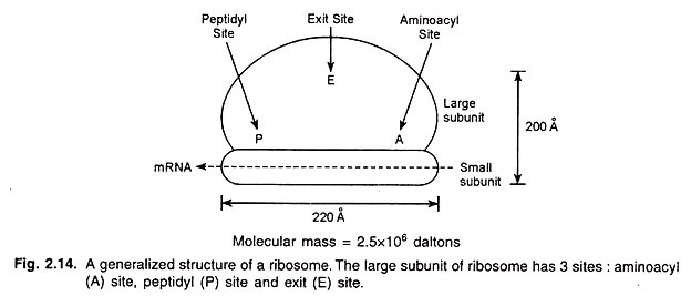
Ribosomes are 200-250 A in diameter; they are regarded as moving factories for protein synthesis. Each cell has a large number of ribosomes e.g., a growing bacterial cell contains about 20,000 ribosomes per genome. In prokaryotic cells, ribosomes are free in the cytoplasm but in eukaryotes, they are mostly found attached to the ER and the outer membrane of the nuclear envelope.
Each ribosome consists of two subunits, called large and small subunits (Fig 2.14). In Escherichia coli, one 50S subunit and one 30S subunit join to form a 70S subunit together give rise to an 80S ribosome. The ribosomes are composed of proteins and RNA called ribosomal RNA or rRNA (Table 2.4).
In bacteria, rRNA accounts for about 80% of the total cellular RNA. In eukaryotes also, rRNA constitutes a major part of the cellular RNA.
A ribosome has three transfer RNA (tRNA) binding sites:
(1) “A” (aminoacyl) site, (2) “P” (peptidyl site), and (3) “E” (exit) site. A tRNA carrying an amino acid enters the A site and then moves to P site. The transfer RNA leaves the ribosome through the E site. During protein synthesis, messenger RNA (mRNA) binds to the ribosome. A ribosome has enough space to bind 40 bases of the mRNA.
Organelle # 10. Chloroplast:
In the cells of green plants, photosynthesis occurs in a specific organelle called chloroplast.
In 1905, Blackman discovered that photosynthesis occurs in two major steps:
(1) Light reaction which is responsible for absorbing light and producing O2, and
(2) Dark reaction in which the energy stored in NADPH (nicotinamide adenine dinucleotide phosphate) and ATP (adenosine triphosphate) produced during light reaction is utilized for the reduction of CO2.
Organelle # 11. Mitochondria:
In 1850, Kolliker described the presence of specific particles in the cytoplasm of muscle cells. This was called by various names, but mitochondrion (thread-like granules) coined by Benda in 1897 is universally accepted.
Since the mitochondrion (pleural = mitochondria) is associated with energy producing pathways, viz. Krebs cycle, electron transfer and oxidative phosphorylation, it is regarded as the “power house” of the cell.
Mitochondria appear as ellipsoid or oval structures in thin sections and range from 3 to 10 p m in length and 0.2 to 1 p m in diameter. The number of mitochondria per cell varies form several hundred to few thousands. In thin section electron micrographs of cells, many mitochondria are interconnected with each other. They contain proteins, lipids, RNA and DNA.


