This article provides information about Chromosomes; its Parts, Functions and Types:
Different Parts of Chromosome:
(1) Pellicle and matrix
(2) Chromonemata
(3) Chromomeres
(4) Centromere
(5) Secondary constrictions
(6) Satellite bodies
Contents
[I] Pellicle and matrix:
Each chromosome is bounded by a membrane called pellicle. It is very thin and is formed of achromatic substance. This membrane encloses a jelly-like substance which is usually called matrix. In the matrix is present the chromonemata. The matrix is also formed of achromatic or nongenic material.
The presence of matrix is controversial but in one organism, Luzula compestris (Woodrush), a plant belonging to the family Juncaceae, its existence is clearly indicated. Recently by the electron microscopic study of the chromosome the presence of pellicle and matrix between the coils of chromonema has been rejected (Darlington, 1937; Ris, 1945).
The function and structure of the matrix is not known. Presumably it aids in keeping the chromonemata with in bounds so that the manoeuvres of the chromosome during cell division can take place unhindered. It may also serve as an insulating sheath for the genes during cell division.
[II] Chromonemata:
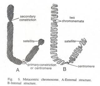
Within the matrix of each chromosome are found embedded two identical, spirally coiled threads, the chromonemata or chromonematal fibrils. Both the chromonemata are so tightly coiled that they appear as a single thread of about 800 A thickness.
This condition is found during prophase and sometimes during interphase. Chromonema was first of all observed by Baranetzky in 1880, in the pollen mother cell of Tradescantia, and was called chromonema (singular) by Vejdovsky in 1912.
At metaphase each chromosome consists of two symmetrical structures, the chromatids, each of which contains a single DNA molecule. The chromatids are attached to each other only by the centromere and become separated at the start of anaphase, when sister chromatids migrate to opposite poles.
Anaphase chromosomes thus have only one chromatid, whereas metaphase chromosomes have two chromatids. Chromatids are the functional unit of the chromosome in cell division, in gene segregation and in crossing over. Chromonema is the gene-bearing portion of chromosome, although each chromonema may contain non-genic materials that serve to maintain its integrity.
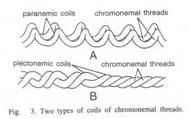
The chromonema may be composed of 2, 4 or more fibrils depending upon the species. This number of fibrils in the chromonema may depend on the different phases since at one phase it may contain one fibril and at other phase it may contain three or four fibrils. These fibrils of the chromonema are tightly coiled with each other. The coils are of two types. —
1. Paranemic coils:
When the chromonemal fibrils are easily separable from each other, such coils are called paranemic coils.
2. Plectonemic coils:
Here the chromonemal fibrils are closely intertwined and they cannot be separated easily. Such coils are called plectonemic coils.
The degree of coiling of the chromonemal fibrils during cell division depends on the length of the chromosome. There are three types of coils: (i) Major coils of the chromonema possess 10-30 gyres; (ii) Minor coils of the chromonema are perpendicular to the major coils and have numerous gyres as observed in meiotic chromosomes. If splitting has not yet occurred at this stage, there will be a single chromonemata. (iii) Standard or somatic coils are found in the chromonema of mitosis where chromonemata possess helical structures, resembling the major coils of the meiotic chromosome.
[III] Chromomeres:
It was demonstrated that the chromosomes particularly in early meiotic prophase, were differentiated into morphologically distinct regions which were constant in size and position. These regions called chromomeres, were first described by Balbiani in 1876 and by Pfitzner in 1881.
Chromomeres are present as beadlike bodies over the chromonemata and the regions in between are designated as inler-chromomeres. Chromomeres structurally different from the remainder of the chromonemata because of a “distinct reactivity of its own in nucleic acid synthesis.”
In other words, chromomere is of great size than adjacent portions of the chromonema because of its ability to synthesize or accumulate on itself, greater amounts of stainable nucleic acid or nucleoprotein. Chromomeres are regions of the superimposed coils confirmed by the electron microscopic observations. These chromomeres are considered to carry the genes during inheritance.
Belling (1928) found about 1,500 to 2,500 independent chromomeres in haploid karyotype of Lilium and Aloe. They are clear in salivary gland chromosomes and ‘lamp brush’ chromosomes of oocytic nuclei. Ultimately, each chromomere corresponds to a single genetic locus.
[IV] Centromere or primary constriction:
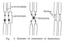
A lighter staining region appears as a constriction or thinner segment of the chromosome and is usually called centromere or primary constriction or kinetochore. It is less densely coiled and thereby slender segment near which the chromatids are held together during metaphase.
The centromere is essential for movement and acentric fragments, which lack a centromere, are therefore incapable of movement on the spindle because spindle fibres only attach to centromeres. Primary constriction or centromere shows positive—Feulgen reaction indicating the presence of DNA.
Under electron microscope, the centromere appears as a disc-shaped protein structure plastered upon the primary constriction. It is about 0.20 to 0.25µ in diameter and is formed of some non-chromatin material.
In cross section it consists of: (i) Electron dense layer 30 to 40 nm thick with convex outer surface. Microtubules of spindle fibres are attached to it and penetrate through it reaching the chromatin fibres, (ii) Inner less dense layer 15 to 30 nm thick lying between electron dense layer and underlying chromatin fibres, (iii) Outer fibrillar material on the convex surface of centromere forming a kind of electron dense corona.
[V] Secondary constriction:
Secondary constrictions mark the locations at which the nucleoli are assembled. The chromosomes in addition to primary constriction (centromere), possess secondary constriction at any point of the chromosome. It is often associated with the nucleolus during interphase and may take part in the reorganization of the nucleolus at the end of cell division.
For this reason a secondary constriction may also be called a nucleolus- organizing region. It appears as a light staining (heterochromatic) region with an additional segment of the chromosome extending beyond it. This extension is satellite body. In a nucleus only two chromosomes usually possess such a zone and hence they are called the nucleolar chromosomes.
The secondary constrictions possess a spiral structure similar to that of the remainder of the chromosome, but the chromatin contained there in exhibits a negative heteropycnosis (Resende, 1940, and Therman-Suomaleinen, 1949).
[VI] Satellite bodies:
The part of the chromosome which is present beyond the secondary constriction is called satellite body or trabant. It varies in size according to the position of the secondary constriction. If secondary constriction is very close to an end of the chromosome, the satellite may be a baiely perceptible dot.
The satellite body as shown in Fig. 6-6 is often seen to be connected to the main body of the chromosome by very light staining strands. Chromosomes bearing satellites or trabants are called SAT-chromosomes (sine acido thymonucleinico). Ordinarily one chromosome in each genome possesses this satellite body and in some cases there may be two or more satellite bodies in a single chromosome.
The extremities or tips of chromosomes are usually called telomeres, a term coined by Muller (1938) to indicate the uniqueness of this portion of the chromosome. The intact end of a chromosome does not enter into permanent association with other parts of the chromosome. The loss of the telomere gives instability to the chromosome to which it was once attached.
This instability results due to the freshly broken end of a chromosome is in an unsaturated state, i.e., it will unite with other similarly broken ends of other chromosomes, or, if the chromosome is longitudinally double, the two broken ends of the chromosomes will unite with each other. Broken ends of chromosomes can sometime ‘heal’ to assume the behaviour and structure of a telomere.
But all experimental evidence on broken chromosomes indicates that this phenomenon is relatively rare. When chromosomes are broken by X-rays, the free ends without telomeres become sticky and fuse with other broken chromosomes. They will not, however, fuse with a normal telomere. Telomeres contain the ends of the long linear DNA molecule contained in each chromatid.
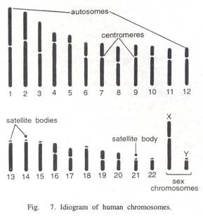
The general morphology of a set of chromosomes or karyotype, of an individual depends upon the dimensions of the chromosomes, position of the kinetochores, (centromeres) and the presence of secondary constrictions and satellite bodies.
A diagrammatic representation of a karyotype showing all of the morphological features of the chromosomes is called an idiogram. The idiogram of man has revealed that five pairs numbered 13, 14, 15, 21 and 22 have nucleolus-organizing regions and satellites.
In general, a particular Karyotype can be designated as representative of the species, and in some instances even of the genus.
Functions of centromere:
(i) Microtubules of chromosomal spindle fibres are attached with the centromere. It helps in chromosomal movement during cell division.
(ii) Centromere is the centre for polymerization of tubulin, a protein used in the formation of microtubules. Thus, it helps in the formation of spindle fibres during metaphase division.
The chromonemata are connected with the kinosome of the centromere. Usually the chromosomes are monocentric, i.e., having one centromere, but some are dicentric (two centromeres) or polycentric. Some species of hemipteran and heteropteran insects have diffuse centromeres, with microtubules attached along the length of the chromosome, such chromosomes are called holocentric.
Acentric chromosomes result due to abnormalities (induced for example by X-ray) in chromosomes. Such chromosomes may break and fuse with other ones. These aberrations are unstable and cannot attach to the mitotic spindle and remain in the cytoplasm.
Most centromeres exhibit a basic structure including one or more chromomeres or kinosomes (dark-staining granules) of varying sizes and inter-chromomeral fibrillae of chromonema. The number of fibrillae varies in a centromere. The centromere is lighter-staining part due to uncoiling of fibrils and less amount of nucleic acids.
The kinosomes may not be visible in each centromere and there may be only fibrillae. In some, there may be symmetry in the distribution of chromomeres and fibrillae or they may be arranged crosswise. Lime-de-Faria (1955) has cytochemically demonstrated that centromere has DNA. Recently, 5 to 7 chromatin fibres (of 230A diameter) have been observed to pass through centromere.
Types of chromosomes:
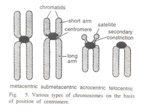
Chromosomes are classified according to their shape, which is determined by the position of the centromere (the point of attachment to the mitotic spindle).
1. Metacentric:
If the centromere is present near about in the middle of the chromosome, then it is called as metacentric, i.e., chromosomes have equal or almost equal arms. Amphibians possess such chromosomes. During anaphase movements, the chromosomes bend at the centromere, so that metacentric chromosomes are V-shaped and acrocentric chromosomes are rod-shaped. Example: Amphibians.
2. Acrocentric:
These chromosomes possess the centromeres near one end forming a long arm and a very short arm or even imperceptible short arm. According to White and Coleman, such chromosomes do not exist in nature. These may be sub-acrocentric and sub-metacentric according to their location in other places. Sub-metacentric chromosomes are L or J-shaped at anaphase, i.e., having arms of unequal length. Example. Mouse.
3. Telocentric:
When centromere is at the terminal or proximal position, then the chromosome is called telocentric. Chromosome is rod-shape at anaphase.
4. Acentric:
If centromere is lacking, the chromosome is termed as acentric.
5. Dicentric and polycentric:
When there are two or more centromeres in each chromosome, then these are referred to as dicentric and polycentric chromosomes, respectively. The two centromeres of dicentric chromosomes tend to migrate to opposite poles, thus leading to chromosomal fragmentation.

