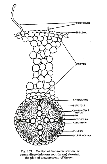
In this article we will discuss about the internal structure of dicot roots with the help of diagrams.
I. Epidermis:
It is single-layered and composed of thin- walled cells. The outer walls of epidermal cells are not cutinised. Many epidermal cells prolong to form long hairy bodies, the typical unicellular hairs of roots. Epidermis of root is also called epiblema or piliferous layer (pilus = hair; ferous—bearing).
II. Cortex:
It is quite large and extensive in roots. Cortex is made of thin-walled living parenchymatous cells with leucoplasts, which convert sugar into starch grains. The last layer of cortex is endodermis. It is of universal occurrence in roots.
Endodermis is composed of one layer of barrel-shaped cells which are closely arranged without having intercellular spaces. The endodermal cells have thickened radial walls, which are called Casparian strips, after the name of Caspary, the gentleman who first noted them.
III. Stele or Central Cylinder:
Next to endodermis there is a single-layered pericycle made up of thin-walled parenchyma cells. Pericycle is the seat of the origin of lateral roots. Vascular bundles are typically radial in roots. Xylem and phloem form separate patches and are intervened by non-conducting cells. In dicotyledonous roots the number of bundles is limited.
Xylem has protoxylem towards circumference abutting on pericycle and metaxylem towards centre. This is called exarch arrangement (of endarch arrangement of stems). Phloem with sieve tubes, etc., form patches arranged alternately with xylem. A small patch of sclerenchyma cells is present outside every group of phloem.
Conjunctive Tissue:
Thin-walled parenchymatous cells lying in between xylem and phloem groups constitute the conjunctive tissue.
Pith:
At the centre there is a small parenchymatous pith. It may be even absent in dicotyledonous roots.
