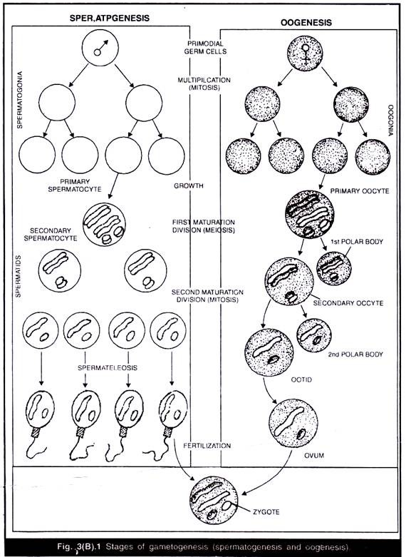
The origin and development of gametes is called gametogenesis (Fig. 3(B).1).
This may be divided into spermatogenesis and oogenesis. Spermatogenesis deals with the development of male sex-cells called sperms in the male gonad or testis.
Oogenesis is the development of female sex-cells called ova or eggs in the female gonad or ovary.
1. Spermatogenesis:
The entire process of spermatogenesis can be divided into following two phases:
(A) Formation of Spermatid:
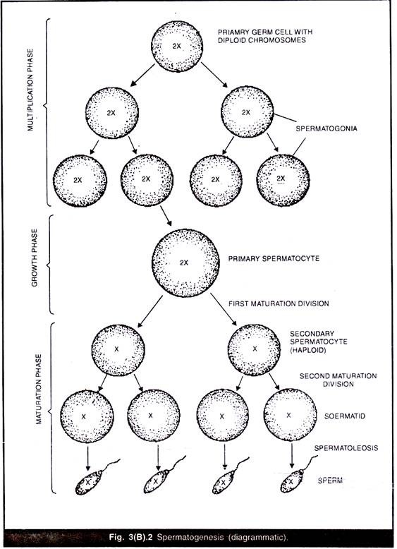
The male gonad known as testis is the site of spermatogenesis. In each vertebrate a pair of testes remains attached to dorsal body wall by a connective tissue called mesorchium. Each testis is formed of thousands of minute elongated and coiled tubules called seminiferous tubules. The inner lining of seminiferous tubules is called as germinal epithelium and is made of primordial germ cells (Primary germ cells) as well as some supporting nutritive cells. The primordial germ cells give rise to spermatids through the following steps (Fig. 3(B).2).
1. Multiplication Phase:
The primary germ cells multiply by repeated mitotic division. The cells produced after the final mitotic divisions are known as spermatogonia or sperm mother cells.
2. Growth Phase:
The spermatogonia do not divide for sometime but increase in size by accumulating nutritive materials from the supporting cells. In mammals such supporting cells are called cells of Sertoli. The enlarged spermatogonia are now called primary spermatocytes.
3. Maturation Phase:
During the phase of maturation, the primary spermatocytes divide by meiosis consisting of two successive divisions. The first division is reductional or disjunctional reducing the chromosome number from ‘2n’ to ‘n’. These cells are celled secondary spermatocytes. Second division is equational resulting in formation of four daughter cells called spermatids.
(B) Spermiogenesis (Spermatoleosis):
This is the second phase of spermatogenesis during which the spermatids produced at the end of first phase are metamorphosed into sperm cells. The spermatid is a typical cell containing a nucleus and cytoplasmic organelles such as mitochondria, golgi bodies, centriole etc, but the nucleus only contains haploid number of chromosomes.
During spermiogenesis or spermatoleosis the following transformations occur in the spermatids:
1. The large spherical nucleus becomes smaller by losing water and usually changes its shape into elongated structure.
2. The Golgi bodies condense into a cap called acrosome in front of the nucleus.
3. Nucleus and the acrosome combinedly form the head of the developing sperm while the cytoplasm with mitochondria and centrioles move downwards and form the cylindrical middle piece behind the head (Fig. 3(B).3).
4. The two centrioles of middle piece develop axial filaments which are bunched into a single thread and extend behind in the form of a long vibratile tail. Thus, spermatid is transformed into a motile sperm divisible into head, middle piece and tail.
2. Oogenesis:
It occurs in the ovary of female animals. It is comparable to spermatogenesis so far as nuclear changes are concerned. But the cytoplasmic specialization in oogenesis is different from spermatogenesis.
It is divisible into following three phases:
1. Multiplication Phase:
The primary germinal cells of the ovary with diploid number of chromosomes (2n) divide several times mitotically so as to form a large number of daughter cells known as oogonia (Fig. 3(B) .4).
2. Growth Phase:
The oogonium does not divide but increases in size enormously to form a primary oocyte. The growth is associated with both nuclear and cytoplasmic growth. The nuclear growth is due to accumulation of large amount of nuclear sap and is termed as germinal vesicle. The cytoplasmic growth is associated with increase in number of mitochondria, endoplasmic reticulum and Golgi complex and accumulation of reserve food material called yolk or vitellin.
3. Maturation phase:
The primary oocyte undergoes two successive divisions by meiosis. The first division is meiosis-I and two unequal daughter cells are produced. The large cell is called secondary oocyte containing haploid (n) set of chromosomes (due to reductional or disjunctional division) and entire amount of cytoplasm. The smaller cell is called first polar body or polocyte containing ‘n’ number of chromosomes and practically no cytoplasm.
The secondary oocyte and first polar body then undergo second maturation division by meiosis-II which is an equational division. As a result of this division one large ovum is formed containing entire amount of cytoplasm and ‘n’ number of chromosomes and a second polar body like the first polar body.
Simultaneously, the first polar body may divide into two polar bodies or may not divide at all. Thus only one functional ovum is formed and the two or three polar bodies soon degenerate. In vertebrates the first polar body is formed after the primary oocyte is released from ovary and has entered into the oviduct. The second polar body is formed only when the sperm enters into ovum during fertilization.
(C) Ripening of Egg:
Oogenesis is followed by the formation of protective coverings called egg membranes. Primary membrane is formed surrounding the plasma membrane of ovum and is secreted by the ovum itself. It is called vitelline membrane in frog and zona pellucida in rabbit. The secondary membrane called chorion is formed from ovarian follicle cells. The tertiary membranes are secreted in oviduct when the ovum passes from ovary to outside. The egg white (albumin), calcareous shell etc. come under this category (Fig. 3(B).5).
The ripe ovum is spherical or oval and non-motile. Depending upon the amount of yolk, it may be as small as 0.15 mm as in mammals (microlecithal); it may be 2 mm as in frog (mesolecithal) or it may be as large as 30 mm as in hen (megalecithal).
In a ripe ovum, the polarity is fixed. The top-most point is animal pole and the bottom point is vegetal pole. The density of yolky cytoplasm increases from the animal pole towards the vegetal pole. In frog, the animal hemisphere is highly pigmented and appears black while the vegetal hemisphere is highly pigmented and appears white.
Significance:
1. The process leads to formation of germ cells or gametes.
2. The normal body cells known as somatic cells are diploid (2n) where as the germ cells are haploid (n).
3. During fertilization one halpoid sperm unites with one haploid ovum to form a normal diploid somatic cell thus keeping the chromosome number constant generation after generation.
4. During first maturation division, the reshuffling of paternal and maternal genes take place resulting in variation.


