The following points highlight the three main components of a Neuron. The components are: 1. Perikaryon 2. Axon 3. Myelin Sheath and Myelinogenesis.
Neuron: Components # 1. Perikaryon:
(a) Its diameter varies from 5µ to 125µ.
(b) It has a cell membrane, cytoplasm, a large vesicular nucleus containing a single prominent nucleolus, numerous rod-like or spherical mitochondria, and a Golgi apparatus.
(c) It also contains certain other organelles which are not present in a usual cell of the body. These organelles include dendrites, presynaptic terminals, sex-chromatin, nissl granules, and neurofribillae etc.
Dendrites:
(a) The cell membrane is projected in the form of a number of filamentous structures known as dendrites.
(b) They may vary from almost none to one metre in length (peripheral sensory nerve fibre).
(c) Each dendron contains nissl granules, mitochondria, and neurofibrillae.
(d) Each dendron on leaving the cells goes on branching repeatedly.
Presynaptic Terminals:
(a) These are small knob like structures which exist on the surfaces of the dendrites and perikaryons.
(b) They are the ends of the nerve fibrils that originate in other neurones next to on which they come to exist.
Sex-chromatin:
(a) It is a large granule often seen adjacent to the nucleolus.
(b) This is found to be present only in females and the sex can be determined by its presence or absence. Hence it is termed as sex-chromatin.
Nissl Granules:
(a) The cytoplasm of a perikaryon shows the presence of basophilic masses known as nissl granules or bodies on the study of special staining techniques.
(b) These granules under electron microscope appear to be composed of many thin membrane bounded cavities or cisternae arranged in a parallel fashion.
(c) Their sizes and number vary with the physiological conditions of the cell, e.g., fatigue, the action of certain poisons. The granular disintegrate into a fine dust which ultimately disappears on the section of the axon.
(d) These granules are similar to the (less numerous) rough surfaced membranes found in the cytoplasm of most cells which collectively make-up the endoplasmic reticulum.
(e) The minute particles consist chiefly of ribose nucleoprotein (RNP) which stain with basic dyes.
Neurofibrillae:
These are aggregation of thin, thread-like structures of variable length and 60-100A in diameter forming a loose network of fibrils in the cytoplasm.
Neuron: Components # 2. Axon:
(a) The axon or nerve fibre arises from the axon-hillock of the perikaryon which is rich in neurofibrillae but has no nissl granules.
(b) It consists of a central core of semifluid axoplasm which flows from the cell body to the axon and not in the reverse direction.
(c) Axoplasm also contains mitochondria, nissl granules, neurofibrillae running parallel to the long axis of the axon. Axoplasm is bounded by a cell membrane known as axolemma which is continuous with the cell membrane of parent cell body.
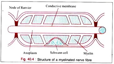
(d) The axon is further surrounded by a sheath known as myelin sheath or medullary sheath which is not present in all the fibres. The fibres having myelin sheath are known as myelinated or medullated nerve fibres and other which have no myelin sheath are known as non-myelinated or non-medullated nerve fibres.
The myelinated nerve fibres are also said to be white nerve fibres on account of the presence of lipids in the myelin sheath. The non-myelinated nerve fibres appear grey for which they are known as grey nerve fibres. All the post-ganglionic fibres of the autonomic nervous system are non-medullated; while all the pre-ganglionic fibres are medullated.
(e) A thin layer of fine reticular fibres surrounding the myelin sheath forms the endoneurium or sheath of Henle.
(f) The axolemma at the terminal end of the axon is also thrown into similar projections—dendrites which come in contact either with the dendrites of the perikaryon of next neuron forming synapses or receptor or effector (e.g., myoneuronal junction).
Neuron: Components # 3. Myelin Sheath and Myelinogenesis:
(a) A specialized set of Schwann cells arranged in a linear manner all around the axolemma forms myelin sheath by a process known as myelinogenesis.
In this process, the Schwann cells grow round the axon and envelop it completely along its entire length so that cell membrane of the Schwann cell can be divided into two parts—one surrounding the axon, i.e., inner part and other outer part which are connected to one another by the double mesaxon.
(b) The Schwann cells start to rotate and go on rotating for several times. As a result, many closely packed, helically arranged layers of double membrane are wrapped around the axon as shown in figure 40.3. This set of membranes is known as myelin sheath.
(c) Each membrane is composed of two layers of lipid substances sandwiched between layers of protein and they form the myelin sheath of the nerve fibre.
(d) The thickness of the myelin sheath is ascertained by the number of membrane layer wrapped round the axon.
(e) The phospholipid sphingomyelin is an excellent insulator that prevents almost all flow of ions outside the nerve fibre.
(f) A small un-insulated area at the junction between each two successive Schwann cells along the axon remains where ions can flow with the case between the extracellular fluid and the axon. The area is said to be the node of Ranvier.
(g) The higher permeability at the nodes of Ranvier causes the impulses to be conducted from node to node by the myelinated nerve fibre rather than continuously along the entire fibre as happen in the un- myelinated nerve fibre. This process is called as salutatory conduction and is helpful in the higher velocity of nerve transmission in myelinated fibres than that in un-myelinated fibres.
(h) The nerve fibres in the central nervous system are myelinated by a different types of cells known as oligodendroglia and by a different process.
(i) The Schwann cells although surround the axon of un-myelinated nerve fibres, they do not spin a myelin sheath around them.
(j) Neurones in different parts of the body differ markedly from one another in the size and shape of the cell body; length, size and number of dendrites; the length and size of the axon; the number of presynaptic terminals which may range from hundred to several thousand.