The following points highlight the two main types of membrane potential. The types are: 1. Resting Membrane Potential 2. Action Potential.
Type # 1. Resting Membrane Potential:
The resting membrane potential may be defined as the net difference in the charge between inside and outside membrane requiring little or no expenditure of energy. In a neurone, this is about -85mV so long as this neurone is in a resting stage and is maintained almost constant.
It is believed to arise:
(1) From the unequal distribution of ionic substances on the two sides of the membrane due to sodium and potassium pumps.
(2) Due to semi- permeability of the membranes.
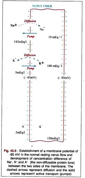
In the resting condition, the concentration of Na+ is about 142 mEq/litre outside the cell and 10 mEq/litre inside the cell; while that of K+ (main diffusible cation) is 5 mEq/litre outside the cell and 140 meq/litre inside the cell due to sodium and potassium pump respectively. There is also a very high concentration of non-diffusible anions, 150 mEq/litre inside the cell in contrast to a very low concentration of only 5 mEq/litre outside the cell.
Simultaneously, K+ is easily diffusible through the membrane; while Na+ and non-diffusible anions diffuse very poorly due to selective permeability of the membrane. Therefore, K+ diffuses from inside to outside of the membrane removing positive (+) charge from the inner side of the membrane while building up positive (+) charge on outer surface of the membrane.
This results in overall excess of negativity on the inner side as compared to the positivity on the outer-side. This comes out to be in between -75 and -95 millivolts with an average of -85 millivolts. This means that the interior of the axon (or nerve cell body) is about 85 mv negative with respect to the exterior in the resting stage of the nerve.
As this potential is dependent on diffusion of K+, it is also called as diffusion electrical potential. There can be another type of electrical potential due to active transport of ions.
Calculation of the Membrane Potential:
The resting membrane potential can be calculated by Nernst equation. When a concentration difference of ions across a membrane causes diffusion of ions through the membrane creating a membrane potential, the magnitude of the potential is determined by the difference in tendency of the ions to diffuse in one direction versus in the other direction.
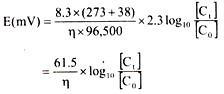
This is determined by the following formula:
![]()
where, E(mv) = Membrane potential in millivolts.
R = Gas constant = 8.3 joules/°C
T = Absolute temperature = 273°C
n = Valency of the diffusible ions
F = Faraday = 96,500 coulombs
Ct = Concentration of diffusible ions inside the cell.
C0 = Concentration of diffusible ions outside the cell.
At a temperature of 38°C (body temperature), this equation results in:
As K+ ions are the main diffusible ions and their Concentration inside the cell is about 30 times as that outside the cell, so the electrical potential created by this concentration difference of K+ ions will be:
![]()
This shows that the calculated value for the K+ diffusion potential (-90 mV) is normally slightly more than the actual measured value of the resting membrane potential (-85 mV). This is due to non-consideration of Na+ and non-diffusible protein anions which also have a slight tendency to diffuse to the interior and exterior of the cell respectively.
Obviously, either of these effects would reduce the potential to slightly below that calculated for potassium diffusion alone.
Type # 2. Action Potential:
So long the membrane of the nerve fibre remains completely undisturbed, the membrane potential remains about -85 mV. This is said to be the resting membrane potential.
But when this membrane is disturbed by a stimulus (e.g., electrical, chemical, mechanical) which can suddenly increase the permeability of this membrane to Na+ then a sequence of rapid changes in the resting membrane potential takes place. This lasts a minute fraction of a second and is followed immediately by return of the membrane potential to its resting value.
This sequence of rapid changes in the electrical potential is said to be the action potential which is discussed below:
It has been found that in the resting condition, the external surface of the membrane is positive with respect to its interior.
When this membrane is suddenly stimulated, the permeability of the membrane to Na+ increases transiently lowering the relative permeability to K’ and, therefore, many of the Na+ rush to the interior of the fibre carrying sufficient positive charge to the interior to cause complete disappearance of the normal resting potential and usually enough charge to develop a positive (+) state inside the fibre instead of the normal negative (-) state.
This positive state inside the nerve fibre is called as the reversal (from negative to positive) potential and this process is said to be the de-polarisation of membrane.
As the positive potential inside the nerve reaches a peak which is known as the peak potential or spike potential, the process of de-polarisation stops and the membrane again becomes al-most completely impermeable to Na+ and relatively more permeable to K+ so that the K+ ion leaves the fibre faster than the Na+ enters the fibre.
Hence, the reversal potential inside the fibre disappears and the normal resting membrane potential returns to the site of stimulation and the fibre once more becomes responsive to further stimulation. This process of restoration of the resting membrane potential from the reversal potential is said to be the re-polarisation of the membrane.
Mechanism of Action Potential:
The exact mechanism responsible for action potential is not yet known.
However, one of the theories to explain it is as follows:
It is postulated that under normal circumstances the sodium “channels” in the membrane are lined with Ca++ repelling the Na+ and other cations. Hence, Ca” resists the passage of Na+ through these sodium channels. Further, when the membrane is stimulated, these Ca++ are dislodged from their binding sites and some Na+ begin to move inwards.
The inward movement of the in-rushing Na+ theoretically dislodge more and more Ca++ from their ending site for which more Na+ rush inward, and the process is continued till no Ca++ remain to resist the passage of Na+ through Na+ channels. The diffusion of the Na+ inside the membrane causes reversal potential with the development of positivity inside the membrane and negativity outside.
This positivity inside the membrane reaching the peak of spike potential opposes further inflow of the positively charged Na+ allowing the Ca++ to begin rebinding with the binding sites along the Na+ channel and the highly concentrated K+ begin moving outwards through the so-called potassium “channels” – probably separate from sodium channels.
As the Ca++ bind, Na+ conductance decreases which allows still more Ca++ to bind and thus another vicious cycle sets in which operates in the opposite direction and continues until the membrane becomes almost totally impermeable to Na+ once again and acquires the similar resting membrane potential.
Propagation of Action Potential:
The electrical stimulation of a nerve fibre initiates at the point of stimulation action potential which in turn excites the adjacent portion of the membrane resulting in the propagation of nerve impulse. Fig. 40.8 shows that when a nerve fibre is stimulated at any point, a sort of current flows inwards through this point and outwards through a part of resting membrane ahead of this, thus completing a circuit.
In some unknown way, the current flowing through resting membrane now increases the membrane’s permeability to sodium which immediately allows Na+ to diffuse inward through the membrane and thus the action potential is developed in this area.
The newly activated area causes local circuits of current flow still further along the membrane causing progressively more and more de-polarisation. Thus, the de-polarisation process propagates in both directions from the point of stimulation along the entire length of the nerve fibre. This transmission of the de-polarisation process along a nerve or muscle fibre is called as the nerve or muscle impulse.

Within few moments the de-polarisation is followed by re-polarisation which also propagates in the same direction in that de-polarisation had previously propagated as shown figure 40. 9: