In this article we will discuss about Mitochondria in Plants and Animals. After reading this essay you will learn about:- 1. Subject-Matter of Mitochondria 2. Morphology of Mitochondria 3. Structural Variations 4. Chemical Composition 5. Development 6. Origin.
Contents:
- Essay on the Subject-Matter of Mitochondria
- Essay on the Morphology of Mitochondria
- Essay on the Structural Variations in Mitochondria
- Essay on the Chemical Composition of Mitochondria
- Essay on the Development of Mitochondria
- Essay on the Origin of Mitochondria
Contents
Essay # Subject-Matter of Mitochondria:
There are several synonymous terms for mitochondria such as chondriochonds, chondriomes, mitosomes, chondriosomes and so on. It was Kolliker who observed granule-like structures in the muscle cells of insects in the year 1880. Flemming (1882) named them ‘fila’ and later on Altmann (1890) named them ‘bioplasts’. In 1897, Benda demonstrated similar objects in cells and assigned the name mitochondria to them.
Meves described mitochondria in plant cell in 1904. The advent of electron microscopy took the study of cell structure to a new level and as early as 1947 Buchholz published some electron micrographs of mitochondria teased out of Tsuga eggs and maize pollen mother cells but little internal details were available from the crude preparations.
The internal structure of mitochondria was described independently by Palade and Sjostrand from ultrathin sections of animal cells in 1953 and the same basic organization has subsequently been shown to hold for all types of mitochondria.
Lewis and Lewis (1914) demonstrated the possibility that mitochondria are concerned with some metabolic activity of the cell and Hogeboom (1948), Kennedy and Lehninger (1948, 1950) and Lehninger (1951) have shown that mitochondria are the chief sites for cellular respiration.
The mitochondria are virtually present in all the aerobic cells. They are absent in bacteria, other prokaryotic cells and mature red blood cells of the multicellular organisms.
The mitochondria are colourless bodies widely distributed in the ground substance of the cytoplasm. These are easily differentiated from other cytoplasmic components by staining process. They are selectively stained by a special stain Janus green.
The mitochondria are of different shapes. They may be fibrillar, spherical, rod shaped and oval and they may change from one form to another depending upon the physiological conditions of the cells. Mitochondria measure usually from 0.2 µ to 2.0 µ in diam. and reach a length up to 40 µ.
Generally, the length of mitochondria ranges from 3 to 5 µ and the size of mitochondrion is greatly influenced by the cell environment (pH, osmotic pressure, etc.). They contain numerous enzymes which take part in the oxidative steps of Kreb’s cycle in respiratory process.
The high energy phosphate compounds such as Adenosine triphosphate (ADP) and Adenosine triphosphate (ATP) are also synthesized and stored in mitochondria. These phosphate compounds after breakdown liberate tremendous amount of energy which is required in the completion of many chemical processes of the cytoplasm.
This is why mitochondria are regarded as ‘power house’ of the cell. They are the principal but not the only sites of oxidation since the oxidation of some compounds also takes place in the ground substance of cytoplasm with the help of enzymes present therein. Mitochondria are found in association with other cytoplasmic organelles in some cells (Fig. 4.1).
Essay # Morphology of Mitochondria:
Light microscopy can reveal no more than overall shape of mitochondria and for their internal structure one has to depend on electron microscopy. The ultrastructure of cell reveals that mitochondria are special type of containers bounded by two membranes of lipoproteins.
The mitochondrial membranes are 50 to 70 Å thick and they can be resolved to display the triple layered unit membrane profile. These membranes are sturdy but flexible allowing contraction and expansion.
The outer membrane is of smooth outline and shows high degree of stretching power. The inner membrane is highly infolded or involute to form plate or tube-like structures, called cristae mitochondriales or mitochondrial cristae, of variable form and number. Except in the regions where it infolds to form cristae, the inner membrane follows the contours of the outer membrane.
The two membranes are separated by an intermediate space or inter-membrane space or outer chamber of fairly constant width which is 20 to 60 A wide and continuous with the intracristal space or lumen. The internal space bounded by inner mitochondrial membrane is called inner chamber. In a number of cell types connections have been observed between the outer and inner chambers (Figs. 4.2 and 4.3).
(i) The Mitochondrial Membranes:
In recent years the two mitochondrial membranes and the chambers they limit have been separated by density gradient method centrifugation. The outer mitochondrial membrane can be separated by causing a swelling and breakage followed by contraction of the inner membrane and matrix.
The outer membrane is much lighter requiring stronger centrifugal force or less-gradient for separation and is more susceptible to fixation damage, sometimes disintegrating while the inner membrane is still intact. The other difference between these membranes lies in their osmotic behaviour.
The inner mitochondrial membrane is an osmotic barrier to substances such as sucrose and contracts and expands in response to changes in sucrose concentration. The outer mitochondrial membrane is permeable to electrolytes, water, sucrose and molecules as large as 10,000 daltons. The inner membrane is normally impermeable to ions, as well as to sucrose.
The inner mitochondrial membrane and matrix both intact when separated from the outer membrane are collectively referred to as mitoplast. The mitoplast possesses pseudopodic processes and is able to carry out oxidative phosphorylation.
The outer membrane has about 40 per cent lipid content as compared to 20 per cent in the inner membrane, contains more cholesterol and is rich in phos photidyl inositol but lower in cardiolipin.
The lipid/protein ratio in outer membrane is quite different from that of inner membrane (0.8 in outer membrane and about 0.3 in the inner membrane). The outer mitochondrial membranes have been found to be pitted in six species of higher plants. The pits measure 25-30 Å in diameter. In animal mitochondria, the outer membranes have not revealed any perforations.
As regards the true width of inter-membrane space, there still exists an uncertainty and, indeed, the very existence of inter-membrane space and the intracristal luman in mitochondria is doubtful as, in fact, different fixatives- and treatments give different results. Both the common fixatives, osmium tetraoxide and potassium permanganate, usually produce a narrow but distinct inter membrane space.
Freeze-etching which is assumed to stabilize organelle morphology unchanged, has shown that the two membranes of yeast mitochondria are closely adpressed leaving no inter-membrane space, while the intracristal spaces are narrow, nearly 50 to 100 Å across.
The mitochondrial space appears very narrow in freeze etched onion (allium cepa) root tip cells. Quick freezing of animal tissues have yielded micrographs showing no gap between the outer and inner membranes and no gap between the membranes of the cristae.
Thus, there may be much less space between the membranes in vivo than that indicated by conventional electron microscopy. The inter-membrane and intracristal spaces have so far revealed no visible structure and appear electron translucent. They are generally assumed to contain an aqueous fluid.
(ii) The Cristae:
The inner mitochondrial membrane limiting the mitoplast has two faces; the outer cytosol or C-face toward the outer chamber of mitochondrion and an inner matrix or M-face toward matrix.
A careful examination of mitochondrial cristae in several groups of plants and animals indicates that there are two types of cristae:
(a) Tubular invaginations (microvilli) and
(b) Plate-like folds (true- cristae). Plant mitochondria are often stated to possess predominantly the tubular or microvillous type of cristae whereas the animal mitochondria usually possess plate- like cristae, but this statement is too sweeping.
Tubular cristae do occur in the majority of algae and fungi but by no means in all. As far as the higher plants are concerned, Opik (1974) is of the opinion that really unequivocally tubular cristae are in minority; some good examples may be found in companion cells of pisum sativum and phaseolus vulgaris. In plants, the mitochondrial cristae are much less regular than well stacked folds in animal mitochondria (Fig. 4.5).
Regular orientation of cristae has, however, been observed in some pollen mitochondria and sometimes in yeast. In plant mitochondria one can find cristae of different shapes and orientations: regular plates, irregular folds, swollen sacs (Fig. 4.6), narrower vesicles, finger like tubules.
Tubular cristae are not uncommon in animal mitochondria. In many Protozoa and steroid synthesizing tissues including the adrenal cortex and corpus luteum the mitochondria possess regularly packed tubular cristae.
There is a direct correlation between the number of cristae and the volume of the matrix. Where there are relatively few cristae, there is much matrix and increase in the number of cristae affects the amount of matrix in the inner compartment of the mitochondrion.
(iii) Mitochondrial Particles:
A critical examination of negatively stained preparation, as well as positive contact preparations has revealed the existence of regular array of particles along the surface of cristae or inner mitochondrial membrane facing the matrix. Discovered first by Fernandez-Moran, they are called inner membrane sub-units or elementary particles or factors of rocker (F1) or oxysomes.
These elementary particles have been observed on the cristae of both plant and animal mitochondria. According to Fernandez-Moran, each of these repeating units is tripartite structure consisting of three parts, a polyhedral or roughly spherical head, a narrow stem or stalk and a roughly cuboid base.
Tripartite unit = base-piece + stalk + head piece.
The stalks (narrow stems) measure nearly 40 to 60 Å in length and 30 to 40 Å in diameter, the heads measure approximately 75 to 110 Å in diameter and bases measure approximately 40 Å. The centre to centre distance between the sub-units measures nearly 110 Å.
The base pieces of these particles form a part of the inner membrane surface (Fig. 4.4). The mitochondrial membranes have about 2,700 particles per square micrometer and depending upon the size and type of mitochondrion, there are from 10,000 to 1,00,000 elementary particles per mitochondrion.
The oxysomes occur in thickness of the inner mitochondrial membrane but they can be seen out of the membrane surface only under special mounting conditions. These particles play an important role in respiration. Functionally, the elementary particles are associated with the presence of enzymes for oxidative phosphorylation and with mitochondrial adenosine triphosphatase.
Green (1959) and Green and Hatefi (1961) have proposed a model for structure of the inner Cristal membrane based on sub-fractionation and electron microscopy. The membrane has been fractionated into the following four functional complexes each with a total molecular weight of approximately 5x 10⁵ and containing enzymes, structural proteins, phospholipids and insoluble cofactors.
Complex I:
NADH-Co Q reductase, catalysing electron transport from NADH to Co. Q.
Complex II:
Succinate-Co Q reductase, catalysing electron transport from succinate to Co-Q.
Complex III:
Co. Q-cytochrome c reductase, catalysing electron transport from Co. Q to cytochrome c.
Complex IV:
Cytochrome c oxidase.
Complexes I, III and IV or II, III and IV in sequence constitute the electron transport chain. CoQ and Cyt. c are considered to act as mobile connecting links or mobile electron carriers between complexes.
The base-pieces which are joined laterally through their phospholipid moieties in the membrane are not detachable from the membrane without destroying it, while the stalks and heads can be detached leaving the membrane still intact. The detachable portion is believed to be identical for all.
Green and Silman (1967) considered electron transfer chain to be located exclusively in the base piece of elementary particles. The stalk contains a protein of low molecular weight (OSCP, oligomycin sensitivity conferring protein) that binds oligomycin and does not show any enzyme activity.
The only enzyme activity detected in the head piece is ATP synthetase, which is probably active in ATP synthesis under normal conditions. The stalk probably acts as a link for couplig electron transport in the base pieces to terminal stages of oxidative phosphorylation (i.e., ATP synthesis) in the head piece.
According to Green, the complete electron transfer chain corresponds in size to the elementary particle. Chance, on the other hand, suggests that each elementary particle carries a single electron carrier component and some additional proteins.
Chance and Parsons (1963) have suggested the term oxysome to represent a factor for coupling phosphorylation to respiration. The elementary particle would then consist of oxysomes or parts thereof.
Matrix:
The inner chamber contains a translucent substance, the mitochondrial matrix. The solid component of the matrix is chiefly protein (about 56%). The matrix presents a finely granular texture or may give impression of a meshwork of fibrils. In the matrix there may occur electron-opaque granules, nearly 60 Å or more in diameter or electron translucent granules in amorphous ground mass.
These granules are now considered to be sites for binding divalent cations, particularly Mg++ and Ca++. Matrix contains a high concentration of all soluble enzymes of Krebs’ cycle and those involved in fatty acid oxidation. Nass and Nass (1963) reported the presence of mt-DNA filaments in mitochondrial matrix of several animal cells and since then DNA filaments have been seen in many plant mitochondria.
Mt-DNA occurs as fine and diffuse filaments or as coagulated opaque mass in electron translucent area of the matrix.
The mt-DNA filaments may be connected to the cristae or inner membrane. In animal, mitochondrial DNA is small, about 5 H long and ring- shaped (Fig. 4.7) like bacterial or phage DNA, but it is linear in plants except yeasts (26 H long). Generally a mitochondrion contains only one mt-DNA containing region, but sometimes several such regions may be observed, especially in elongated mitochondria.
Mitochondrial DNA is different from nuclear DNA in having different purine and pyrimidine bases. Rabinowitch (1968) has shown that mitochondrial DNA contains more Guanine and Cytosine (GC) contents than the nuclear DNA. Mitochondria have a DNA polymerase different from nuclear DNA polymerase.
In mammalian cells mitochondrial DNA constitutes less than 1% of nuclear DNA. It has very low molecular weight (about 106) and contains few coded information. Mitochondria can use their DNA to synthesize complementary RNA.
Like plastids, mitochondria have their separate protein synthesizing machinery, i.e., they have their own t RNAs (transfer RNAs, small sized 55S ribosomes, aminoacids and enzymes necessary for activating aminoacids.
Mitochondrial ribosomes (mitoribosomes) can be seen in the matrix, looking slightly smaller than the cytoplasmic ribosomes (cytoribosomes). They may be found randomly distributed or can form small groups, possibly polysomes. Sometimes mitoribosomes appear associated with cristae or inner membrane.
They can synthesize some, but not all of their constituent proteins. They cannot synthesize cytochrome c, the synthesis which takes place on the ribosomes in extramitochondrial cytoplasm. Mitochondria synthesize structural proteins that are thought to act as self-assembly organizers for the lipids, the enzymes and the other proteins which constitute a complete membrane system.
Parson and Simpson (1967) have shown that the mitochondria contain the enzyme DNA polymerase which helps in the replication of mitochondrial DNA. Mitochondrial DNA is self-duplicating unit and it has been suggested that the duplication of mitochondrial DNA is controlled by nuclear genes. Mitochondria are, therefore, considered as semi-autonomous structures in the cells.
Essay # Structural Variations in Mitochondria:
The structure of mitochondria is not constant in all plants and animals. The mitochondria present several structural variations. These variations are caused due to the differences in number of cristae, size, shape and arrangement of cristae and fusion of mitochondria themselves.
1. Variations due to number of cristae:
The number of cristae per unit volume varies greatly in the mitochondria of most plant and animal cells and the number of cristae seems to be correlated with oxidative activity in mitochondria.
2. Variations due to the structure and orientation of cristae:
Normally cristae in rod shaped mitochondria are oriented perpendicular to the long axis of the mitochondria, but this is too a general statement.
The cristae may be parallel to long axis of the mitochondrion, as in marrow and striated muscles or they may be arranged as vesicles or occasionally branched to form a network of connecting chambers, as in human leucocytes, protozoa, parathyroid gland and com mitochondria, or they may be extremely reduced.
In some mitochondria the cristae may be arranged in concentric rings and in some cases they may be haphazardly arranged.
3. Variations due to fusion:
De Robertis (1957) has reported a peculiar state in spermatids of certain insects in which all mitochondria in the cytoplasm aggregate around the nucleus and they all fuse together to form a single mitochondrial body.
Essay # Chemical Composition of Mitochondria:
The gross chemical composition of mitochondria varies in different cells of both plants and animals. Typically, however, by dry weight mitocondria are about 65 to 75% protein and nearly 25 to 30% lipids. Of the lipid component, 90% is phospholipid and 10% carotenoids, cholesterol, vitamin E and other traces.
Mitochondria contain sulphur, iron, copper and some vitamins in traces which are mostly related to enzyme activities. They also contain small amount of mt-DNA and about 0.5% RNA. When mitochondria are separated from a cellular environment and ruptured, some of the enzymes associated with matrix are released as soluble proteins while the other enzymes remain firmly bound to the membranes.
The mitochondria contain about 70 enzymes and about a dozen co-enzymes and numerous co-factors. Soluble enzymes from mitochondria include all enzymes of the tricarboxylic acid cycle (TCA) except some dehydrogenases, those that catalyze β oxidation of fatty acids and others that catalyze transamination of amino-acids and synthesis of protein.
Membrane bound enzymes of mitochondria are these essential for electron transport chain and oxidative phosphorylation. Mitochondria do not contain the enzymes for anaerobic glycolysis which occurs in the groundplasm.
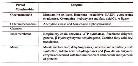
Lehninger (1969) has identified the following enzymes in different regions of mitochondria:
Essay # Development of Mitochondria:
In the meristematic cells of higher plants the mitochondria are not well differentiated and their cristae are irregular, sac-like and a large proportion of matrix is occupied by electron translucent regions with conspicuous DNA filaments. Ribosomes and ribosome like particles can also be seen frequently in them.
The undifferentiated mitochondria undergo a series of changes in the course of maturation and the development takes place in the following steps:
(i) Mitochondrial size may increase.
(ii) Changes in the opacity of matrix and dilation of the cristae take place.
(iii) With the advancement of cell age the DNA filament and the surrounding electron translucent areas become less conspicuous or may become completely obliterated. This does not necessarily mean a disappearance of the mitochondrial DNA which can still be isolated from the mature tissues.
(iv) The mitochondrial number per cell increases as the cells grow; accurate numerical estimations, however, are not easy and only a few have been made. In maize root cap the number of mitochondria per cell increases from 50 in the cap initial to approximately 175 in the mature cells.
Mitochondria do persist in dry dormant seed tissues of higher plants and in spores. The protoplasmic changes which occur during dehydration greatly lower the membrane contrast. Additionally, the number and width of cristae per mitochondria may decrease as seeds dry out, as for examples, in Pea cotyledons and radicle. DNA and granular mitoribosomes may still be discernible.
Essay # Origin of Mitochondria:
There are at least four different views regarding the origin of mitochondria and these are as under:
(i) Autonomous or de novo origin.
(ii) Origin from promitochondria.
(iii) Origin by division or budding of pre-existing mitochondria.
(iv) Nuclear origin.
(v) Prokaryotic origin.
(i) Autonomous or de novo origin of mitochondria:
According to this view, the mitochondria are autonomous organelles with independent hereditary action and leading symbiotic life. The mitochondria are supposed to have originated de novo from the simple building blocks such as aminoacids and lipids.
Some observations are available where mitochondria were completely damaged and it was found that new chondriomes developed de novo. The significance of this observation can be questioned and it is still not clear whether mitochondria are fully autonomous or semi-autonomous bodies.
(ii) Origin from promitocliondria:
Like plastids, the mitochondria also originate from the special type of cytoplasmic precursor bodies which are called the ‘promitochondria’. These bodies do not have cristae and cannot use oxygen. Such promitochondria have been observed in anaerobically grown yeast. The precursor bodies after a few hours of exposure to oxygen develop into normal mitochondria with cristae [Figs. 4.8 A, B and C].
According to Morrison (1966), “The promitochondria might originate from endoplasmic reticulum or plasma membrane”. The process is illustrated in Fig. 4.9. The hypothesis is not supported by direct evidence, hence not valid at present.
(iii) Origin by division and budding of pre-existing mitochondria:
Mitochondria have been found to undergo division and budding [Figs. 4.8 D and E]. It has been observed with time-lapse cinomatography that mitochondria gradually elongate and then fragment into smaller mitochondria. This observation has been verified in neurospora by radioactive tracer technique. Budding of mitochondria may occur in differentiated cells giving rise to mitochondrial initials.
These can then increase in size and develop cristae. As regards the timing of mitochondrial division, very little information’s are available. In shoot meristem of epilobium hirsutum the mitochondrial multiplication occurs during interphase.
(iv) Nuclear origin of mitochondria:
Sometimes the double layered nuclear wall evaginates to form vesicles which when detached from the nuclear wall give rise to promitochondria or protoplastids. In such a development, the mitochondria are independent bodies.
A claim for the evagination of mitochondria from the nuclear membrane in developing egg cells of the fem pteridium aquilinum has been made by bell and muhlethaler (1964) on electron microscopic evidence, but it has been firmly denied by other authors and recent works from Bell’s own laboratory has failed to produce supporting evidence from radioactive labeling experiments.
Mitochondrial initials may also represent derivation from plasma membrane. Robertson (1964) has suggested a scheme whereby mitochondria may arise from invaginations of plasma membrane (Fig. 4.9) or from endoplasmic membrane.
(v) Prokaryotic origin or symbiotic hypothesis:
When Altman (1890) first described bioplasts, the considered them to be Element-organismen evolved from and related to free living forms such as bacteria. According to symbiotic hypothesis mitochondria may be considered as intracellular symbiotic organisms living in association with higher cells.
The homologies between mitochondria and bacteria are numerous and when considered from an evolutionary view-point, they may be more than circumstantial.
The size of a mitochondrion is of the same order or magnitude as that of bacterial cell. Bacteria bear oxidative enzymes in the cell membrane. The protein synthesizing system in mitochondrion is of bacterial type and the mitochondrial DNA region resembles bacterial nucleoplasm and the circular DNA molecules found in yeast and animal mitochondria can be compared with circular DNA molecule found in bacteria.
There are similarities in the localization of respiratory chains. These similarities make the idea of ultimate origin of mitochondria from symbiotic prokaryotic organisms very attractive. The outer membrane of mitochondria which is different from inner membrane but quite similar to endoplasmic reticulum might be derived from a host cell membrane surrounding the microorganisms.


