
In this article we will discuss about Echinococcus Granulosus (Dog Tape Worm).
Morphology of Echinococcus Granulosus (Dog Tape Worm):
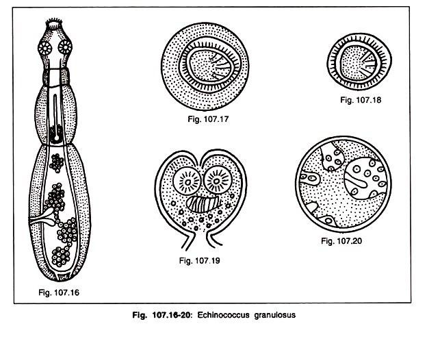
The adult worm (Fig. 107.16) is a minute tape worm, has a scolex, neck and strobila comprising three segments and measures 3-6 mm in length. It has a pyriform scolex provided with four suckers and a protrusible rostellum armed with two circular rows of hooklets. The neck is short and thick. Its egg (Fig. 107.17, 18) cannot be distinguished from that of Taenia and is infective to man, cattle, sheep.
Life cycle of E. Granulosus:
The infected definite host (dog) passes the stool with the eggs onto the ground. While grazing on the polluted ground, the intermediate hosts (sheep, goats and cattle) swallow these eggs, whereas children get infected while playing with dogs.
The eggs hatch in the duodenum, the oncospheres migrate through the intestinal wall, enter the mesenteric venules and become lodged in the capillary filter beds in various organs and tissues (liver, lungs, various organs) and develop into a cystic cavity (hydatid cyst — Fig. 107.19, 20).
When the organs or tissues containing fertile hydatid cysts are ingested by dog, the cysts develop into adult worms. The eggs are passed out in the dog’s faeces.
Clinical features of E. Granulosus:
The damage produced by hydatid cyst of E. granulosus is both mechanical and toxic. The young cysts which develop into embryos lodged in the vital centres may interfere with the function of the organs, damage the organ (brain, orbital capillary, heart valve) and even cause death.
Laboratory diagnosis of E. Granulosus:
Casoni’s test or intradermal test, precipitin test, complement fixation test; haemagglutination; bentonite flocculation test; latex agglutination test; fluorescent test ELISA; exploratory cyst puncture; roentgenogram and recent Computed Tomography (CT) scan and Magnetic Resonance Imaging (MRI) are techniques used for the diagnosis of hydatid disease.
Coagglutination test is very recent sensitive, specific, rapid (within 30 minutes), inexpensive, Dot-blot-ELISA is confirmative test.
Treatment:
Albendazole is most effective than thiobendazole and mebendazole. Surgical technique is also helpful. Combination of praziquantel and albendazole is very effective.
Prophylaxis:
This consists of:
(1) Avoidance of handling infected dogs;
(2) Avoidance of ingestion of raw vegetables polluted with eggs;
(3) Personal hygiene (cleaning hands before eating);
(4) Preventing dogs from eating the carcasses of sheep; cattle and dogs in infected areas; (5) Discarding all infected viscera; in slaughter houses by dumping them into pits inaccessible to dogs and
(6) Educational propaganda in schools.
Polycystic hydatid disease caused by E. vogeli can be treated with albendazole.