In this article we will discuss about the vegetative, asexual and sexual methods of reproduction in lichens.
Vegetative Reproduction:
(a) By Fragmentation.
Death and decay of older parts of the thallus produce smaller pieces which give rise to new thallus. Sometimes the broken pieces (fragments) develop into new thalli, provided they contain both the algal and fungal components.
(b) By Soredia.
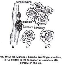
It is the most common method of vegetative reproduction. These are small protuberances, produced on the upper surface by the thallus. They may either occur within definite pustule-like compact structures called soralium (Fig. 10 D) or may arise so abundantly as to spread up like a thin greyish layer of dust. Each soredium consists of a few algae cells surrounded by a mass of hyphae. (Fig. 10 A-C).
Soredia arise from the algal zone below the upper cortex. The cells of the algal zone divide actively and soon get surrounded by the fungal hyphae. Soredia are very light in weight and are easily disseminated by wind or rain wash. After falling on suitable substratum, they develop into a new lichen e.g., Parmelia, Bryoria etc.
(c) By Isidia:
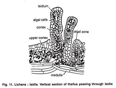
These are the stalked, un-detachable outgrowths produced by the thallus on its upper surface (Fig. 11). Like soredia, the isidia are also composed of both fungal and alga, components but differ from them in being covered with a definite cortex.
The algal component is of the same kind as in thallus. The isidia may be rod shaped (Parmelia sexualities), coralloid (Umblicaria postulata), cigar shaped (Usnea comosa) or scale shaped (collema crispum).
The main function of the isidia is to increase the photosynthetic surface of the thallus. Sometimes these also act as organs of vegetative propagation.
(d) By lobules:
Some dorsiventral outgrowths are produced on the margins of the thallus of Parmelia and Peltigera lichens. These structures are known as lobules and act as organs of vegetative propagation.
Some lichens by forming propagules of different kinds such as phyllidia (leaf or scale like dorsiventral portions e.g., Peltigera), blastidia (yeast like, segmented e.g., Physcia), schizidia (scale like e.g., Parmelia), hormocyst (algae hyphal and fungal hyphae grow together in a chain like manner e.g., Lempholema), goniocysts (unsorallium like structures), etc.
Asexual Reproduction:
By Sporulation:
Certain lichens may also reproduce asexually by means of conidia (e.g., Arthonia), oidia and Pycniospores or pycnidiospores (Fig. 12 A, B). However, it is of rare occurrence. In some cases hyphae break down into small pieces known as oidia while pycniospores are produced within the flask shaped structures known as pycnidia (Fig. 12).
Each pycnidium opens to the surface through a small pore known as ostiole. The pycnidial wall is made up of sterile fungal hyphae. Inside the pycnidia fertile hyphae obstruct sexual spores (pycnidiospores) at their tips. After falling on suitable substratum pycnidiospores germinate and coming in contact with appropriate alga, they develop further into a new lichen.
Sexual Reproduction:
The sexual reproduction in Ascolichens and Basidiolichens is like class Ascomycetes and Basidiomycetes respectively. Ascolichens have been studied in more detail from this point of view. The male reproductive organ is called the spermogonium and the female is known as carpogonium. They develop either on the same hypha or on two different hyphae of the same mycelium.
a. Spermogonium:
The spermogonia are flask shaped structures embedded in the upper surface of the thallus. They open outside by a small pore known as ostiole. The fertile hyphae in the cavity of the spermogonium abstract minute rounded cells at its tip. These male cells are known as spcrmatia. In some species of lichens, however, the pycnidia like structures also function as spermogonia (Fig. 7).
b. Carpogonium:
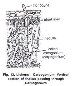
A carpogonium consists of two parts (Fig. 13) i.e., lower coiled multicellular portion called ascogonium and the upper long, Straight, thread like portion called trichogyne. The ascogonium lies deep in the medullary portion while trichogyne emerges out of the thallus and receives spermatia.
Fertilization:
On being disseminated, the spermatia have been found sticking to the protruding tip of trichogyne. This is the only evidence that spermatia function as male gametes. However, Morean and Morean (1928) opined that there is never any fertilization of the protruding trichogyne by spermatia.
After fertilization trichogyne withers. The ascogonium produces freely branched acrogenous hyphae. These hyphae produce asci at their ends. All the structures after fertilization (i.e., developing asci, ascogenous hyphae and ascogonium) are surrounded by the sterile hyphae. It results in the formation of fruiting body which is either a apothecium or perithecium type.
Structure of Apothecium:
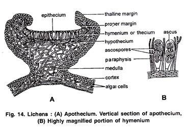
It is round and cup-shaped structure (Fig. 14 A). If the apothecium consists only the fungal component, it is known as lecideine type (e.g., Lecidea, Cladonia, Gyrophora) and if it consists bath algal and fungal components it is known as lecanorine type (e.g., Lecanora, Parmelia).
It can be divided into two parts (Fig. 14 A):
(1) Disc of the Apothecium:
(a) Hymenium (Thecium). It is the upper-most fertile layer of apothecium consisting of a closely packed, palisade like layer of sac-like asci and sterile hair like fungal hyphae known as paraphyses. This layer is also called hymenial layer or hymenium. Each ascus contains 8 ascospores (Fig. 14 B). Ascospores are of various shapes and size, multicellular uni- or bitunicate and uni- or multinucleate.
The sterile tissue that separates the asci is called hamathecium. As many as four types of hamathecial tissues are identified in an ascocarp.
These are:
1. Paraphyses:
Arise from the base of ascocarp and grow upwards (Fig. 15 A).
2. Paraphysoides:
They are formed by stretching of the tissues of ascocarp before the development of asci (Fig. 15 B).
3. Periphysoides:
Arise from the roof of ascoscarp and grow downwards (Eig. 15 C).
4. Periphyses:
Arise in the ostiolar canal and protrude outside the ostiole (Fig. 15 D).
(b) Sub-hymenium:
The region consists of the closely interwoven sterile hyphae. It is present just below the fertile layer.
(2) Margin of Apothecium:
This part surrounds the disc and also forms the edge of the apothecium.
Germination of ascospores:
The ascospores may be simple or septate. They are very light in weight and easily disseminated to a long distance by wind. After falling on suitable substratum it germinates and produces fungal hyphae. The hypha grows into a new lichen thallus, if it comes in contact with an appropriate algal component.
