In this article we will discuss about Neanthes Virens:- 1. Habit, Habitat and Distribution of Neanthes Virens 2. External Features of Neanthes Virens 3. Locomotion 4. Body Wall 5. Coelom 6. Digestive System 7. Respiration 8. Blood Vascular System 9. Excretory System 10. Nervous System 11. Sense Organs 12. Reproductive System and Others.
Contents:
- Habit, Habitat and Distribution of Neanthes Virens
- External Features of Neanthes Virens
- Locomotion of Neanthes Virens
- Body Wall of Neanthes Virens
- Coelom of Neanthes Virens
- Digestive System of Neanthes Virens
- Respiration of Neanthes Virens
- Blood Vascular System of Neanthes Virens
- Excretory System of Neanthes Virens
- Nervous System of Neanthes Virens
- Sense Organs of Neanthes Virens
- Reproductive System of Neanthes Virens
- Swarming of Neanthes Virens
- Fertilisation of Neanthes Virens
- Development of Neanthes Virens
Contents
- 1. Habit, Habitat and Distribution of Neanthes Virens:
- 2. External Features of Neanthes Virens:
- 3. Locomotion of Neanthes Virens:
- 4. Body Wall of Neanthes Virens:
- 5. Coelom of Neanthes Virens:
- 6. Digestive System of Neanthes Virens:
- 7. Respiration in Neanthes Virens:
- 8. Blood Vascular System in Neanthes Virens:
- 9. Excretory Organs of Neanthes Virens:
- 10. Nervous System of Neanthes Virens:
- 11. Sense Organs of Neanthes Virens:
- 12. Reproductive System of Neanthes Virens:
- 13. Swarming of Neanthes Virens:
- 14. Fertilisation of Neanthes Virens:
- 15. Development of Neanthes Virens:
1. Habit, Habitat and Distribution of Neanthes Virens:
Neanthes Virens is found on the sea- shore in the shallow water in rock crevices, hidden under the stones or sea weeds. Some live in tubular U-shaped burrows lined by mucus in sand or mud at tide level. It is carnivorous as it feeds on small insects, mollusks and worms. It is very active during night and passive during the day i.e.; nocturnal.
During night they come out from their hidden place with their heads protruding foe feeding. When breeding periods approaches it leaves the burrow and comes at the water surface to lead a pelagic life, then it is called heteronereis.
Distribution of Neanthes:
Neanthes is cosmopolitan in distribution.It is found abundantly in Europe, North Pacific, America, Alaska and other place.
2. External Features of Neanthes Virens:
(i) Shape and Size:
The body is long, narrow, slender , bilaterally symmetrical , tapering posteriorly and relatively broad anteriorly. It is approximately dorsoventrally flattened with rounded or convex dorsal surface and flat ventral surface surface. The size varies from species to species; it may range from 30 to 40 and 2 to 6 mm in width.
(ii) Colouration:
The colouratiuon of body varies in different species, and it may also vary even in the individuals of same species of different age and sexual maturity. However, N.plelagica is reddish brown. N. cultrifera is greenish N. limicola is brownish in colour, while N, virens is bluish- green in colour having orange or red tinge on the appendages
(iii) Segmentation:
The anterior end of Neanthes Virens is differentiated into a distinct head and the rest of the body is divided by a series of ring-like narrow grooves into a series of segments or metameres or somites arranged in a linear series. All the metameres are nearly alike except the last one which is rounded.
The number of metameres varies from species to species. In N. dumerilli and N. cultrifera, the number of segments is nearly 80, while in N. virens the number of metameres reaches up to 200.
(iv) Division of Body:
The body of Neanthes Virens is divisible into three well marked regions, viz., head, trunk, and pygidium.
1. Head:
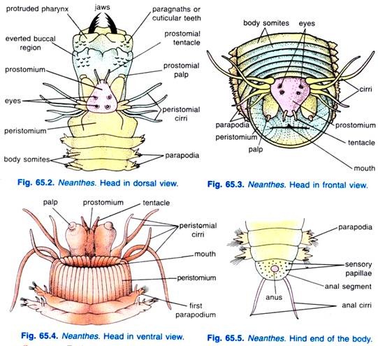
The anterior end of the body possesses a distinct, prominent and well developed head which consists of two main parts, prostomium and peristomium.
(i) Prostomium. (Gr., pro = before; stoma = mouth):
Prostomium is an anterior narrow, nearly triangular fleshy outgrowth. It is situated mid- dorsally in front of the mouth. It is not the true segment of the body as it is derived from pre-oral lobe of the larva.
(ii) Peristomium. (Gr., peri = around; stoma = mouth):
Peristomium is a large ring-like structure carrying ventrally the transverse mouth. The peristomium is formed by the fusion of first two body segments, it forms the lateral and ventral margins of the mouth.
Sensory appendages and organs of head:
Various sensory appendages and organs are found on the head of Neanthes.
These are as follows:
(i) Prostomial eyes:
Prostomium bears four simple, black and rounded eyes on its dorsal surface. These are sensitive to light.
(ii) Prostomial tentacles:
There is a pair of short cylindrical prostomial tentacles projecting forward from the anterior border of the prostomium. These are probably tactile in function.
(iii) Prostomial palps:
These are also two in number but somewhat stout and longer. These are two jointed appendages present on the ventral side of the prostomium but posterior to tentacles. These are also supposed to be tactile organs.
(iv) Nuchal organs:
These are ciliated pits of doubtful nature lying one on each side of the prostomium.
(v) Peristomial cirri:
These are long slender structures present on the anterior side of the peristomium. Four peristomial cirri, two dorsal and two ventral are present on each lateral side of the peristomium. These are tactile organs. These are probably homologous with the parapodial cirri of other body segments.
2. Trunk:
Posterior to the head, the rest of the body which is metamerically segmented (having 80-120 segments) is called trunk. The segments are known as metameres or somites which bear a pair of parapodia are nearly alike except the last.
3. Pygidium:
The last segment of the body called variously as tail, anal segment or pygidium, is elongated, swollen and bears a terminal anus. It bears no parapodia but has a pair of elongated anal cirri which are ventral cirri and several minute sensory papillae. The pygidium represents the posterior part of the larva, and the body segments are formed in front of it.
(v) Parapodia (Gr. para = side + podus = foot):
Each segment of the body, except the peristomium and the anal segment, bears on either lateral side a flat, fleshy, hollow and vertical flap-like out-growths, the parapodia. Huxley first of all gave the name parapodia to these fleshy outgrowths.
Structure:
Each parapodium is a biramous structure consisting of two parts-an upper or dorsal blade, the notopodium and a lower or ventral blade, the neuropodium. Each of these is further divided into two lobes, an upper and a lower. The dorsal margin of notopodium is produced into a short, cylindrical, tactile appendage, the dorsal cirrus and a similar structure is produced at the ventral margin of the neuropodium, the ventral cirrus. Of the two cirri, the dorsal one is much larger than the ventral.
Both the notopodium and neuropodium have a bundle of bristle-like setae or chaetae lodged in a sac formed by the invagination of the epidermis, the setigerous or chaetigerous sac. The setae are capable of being protruded or retracted and turned in various directions by strands of muscular fibres present in the interior of parapodium. Each seta of the bundle originates from a single large formative cell present at the base of setigerous sac.
In the middle of each bundle of setae and deeply embedded in the parapodium is found stout, straight, thick and dark coloured chitinous rod the aciculum which projects only a short distance but does not project beyond the outer edge or the parapodium. At its inner end the aciculum has attached muscles by which protrusion and retraction of the parapodium occurs. The two acicula constitute the endoskeleton of parapodium and serve to support and for attachment of the setal muscles.
The parapodia are the largest in the mid-region of the body and decrease in size towards the anterior and posterior ends of the body. The first two parts of parapodia have no notopodial setae. The parapodia are highly muscular, well vascularised and glandular structures.
Functions:
The parapodia are primarily the organs of locomotion, used both in creeping and in swimming. Since the parapodia are highly vascularized, hence, they also serve the function of respiration.
Setae or Chaetae:
The setae or chaetae are fine but stiff chitionous rods which help in the locomotion by acting as minute paddles; these are protective used for offense and defence and they also provide hold on the smooth surface of the burrow. Each seta consists of two movable articulated parts—the basal shaft or stalk and the distal blade which can be retracted into the shaft.
There are three types of setae, viz.,
(i) Having a small slender shaft and the terminal long blade which is nearly straight, pointed and serrated at one edge,
(ii) Having a large stout shaft and short and slightly hooked blade and
(iii) Having the oar-shaped blade.
Nephridiopores:
In each segment on the ventral side of the body near the bases of Parapodia are found laterally a pair of minute openings, the excretory opening or nephridiopores by which excretory materials are removed.
3. Locomotion of Neanthes Virens:
Locomotion is brought about by the combined action of Para podia, body musculature and to some extent the coelomic fluid. Neanthes Virens shows generally two types of locomotion, viz., slow creeping and fast creeping but sometimes it swims in water.
(i) Slow Creeping:
Slow creeping movements of Neanthes Virens are carried out by the action of Para podia only. During locomotion each Para podium performs two strokes an effective or back stroke and recovery or forward stroke. In the effective stroke, the aciculum of Para podium is extended so that the Para podium is lowered to come in contact with the substratum and moves backwards against the substratum.
In the recovery stroke, the aciculum is retracted so that the Para podium is lifted above and moves forward. The combined effective and recovery strokes of numerous Para podia propel the worm forward. The Para podia of the two sides work alternately causing successive waves along each side of the worm.
(ii) Fast Crawling:
In addition to the parapodial locomotion, undulatory movements of the body cause the worm to crawl or swim rapidly.
Body undulations are caused by waves of contractions in the longitudinal muscles of the body wall. These contractions coincide with the alternating waves of Para podia on the two sides. The longitudinal muscles of one side contract when the Para podia of that side are moved, the muscles relax when Para podia sweep backwards.
4. Body Wall of Neanthes Virens:
The body wall is thick and consists of the following layers:
(i) Cuticle:
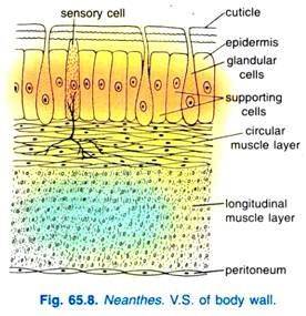
It is thin, tough, chitinous, and non-cellular layer covering the body externally. It is perforated by numerous pores, the openings of epidermal gland cells and exhibits an iridescent lustre due to the presence of two intersecting systems of fine lines.
(ii) Epidermis or Hypodermis:
Below the cuticle and resting on a thin basement membrane is a single cell thick layer, the epidermis which is composed of columnar epithelial cells of which some are glandular and open externally by pores, some are sensory, while others are more or less un-specialised columnar cells or supporting cells.
The epidermis is thick and glandular ventrally particularly near the bases of the Para podia because the glands are larger and more numerous in this region in comparison to the dorsal epidermis. The gland cells produce mucus which lines the burrows of the animal. The epidermis is richly vascularized and helps in respiration.
(iii) Musculature:
Below the epidermis are present the muscles which are of three types, viz., circular, longitudinal and oblique. Circular muscles lie just below the epidermis and form a thin continuous layer around the body. Contraction of these muscles makes the body longer and thinner. Below the circular muscles are present the longitudinal muscles which are better developed than the circular muscles.
They do not form any continuous layer around the body but are arranged in four separate longitudinal bundles—two dorsolateral and two ventrolateral. Contraction of these muscles makes the body shorter and thicker. There are two pairs of oblique muscles in each segment which originate from the median ventral line and pass dorsolaterally to be inserted into the circular muscles on the base of Para podium.
Each oblique muscle is made up of two bundles of muscle fibres, one bundle goes to the dorsal part of the base of Para podium and the other to the ventral part.
The function of these muscles is to retract the Para podium. Protrusion of Para podium is largely due to the pressure of coelomic fluid. But each Para podium has other muscles also, the largest parapodial muscles arise from the circular muscles of the body wall and are joined to the acicula, they extend the acicula and Para podium. All muscle layers are syncytial tissue.
(iv) Peritoneum:
A parietal peritoneum is the innermost layer of the body wall, which lines the musculature internally and forms the lining of coelom; it secretes coelomic fluid.
5. Coelom of Neanthes Virens:
The coelom is an extensive perivisceral cavity having an outer parietal peritoneum and an inner visceral peritoneum which encloses the alimentary canal. The coelom is schizocoelic in annelids having been formed by splitting of the mesoderm into two layers.
Coelom is divided into a linear series of compartments by inter-segmental septa which pass inwards from the body wall but do not quite join the alimentary canal, the septa also have apertures, hence, the coelomic compartments communicate.
Each septum has a double layer of coelomic epithelium containing muscles and connective tissue. The coelom is filled with a coelomic fluid containing amoeboid corpuscles or coelomocytes, and during the breeding season reproductive cells in various stages of development. The coelom communicates with the outside by nephridia and coelomoducts.
Functions of Coelomic Fluid:
The functions of coelomic fluid may be counted as under:
1. It provides turgidity to the body which aids in locomotion.
2. It helps in the protection of the body by absorbing external shocks, if any, and acts as a hydraulic skeleton.
3. It helps in the distribution of various nutritive materials and respiratory gases to the whole body.
4. It helps in removing excretory wastes from the body.
6. Digestive System of Neanthes Virens:
Alimentary Canal of Neanthes Virens:
The alimentary canal is a straight and un-branched tube from mouth to anus. It has two openings to the exterior, an anterior one, the mouth by which food is ingested and a posterior one, the anus through which undigested food remains are removed. The alimentary canal is differentiated into three regions on the basis of its lining-stomodaeum or foregut, mesenteron or midgut and proctodaeum or hindgut.
1. Stomodaeum or Foregut:
It is the anterior region of alimentary canal, lined internally by ectoderm and cuticle. It consists of buccal cavity and pharynx.
(i) Mouth:
The mouth is a transverse slit, lies ventral to the prostomium. It is bordered laterally and ventrally by the peristomium which forms a buccal ring. The mouth opens into a wide chamber, the buccal cavity.
(ii) Buccal Cavity and Pharynx:
The buccal cavity is a short but wide chamber situated inside the peristomium. The buccal cavity leads into a muscular protrusible pharynx extending up to fourth segment. The buccal cavity and pharynx are bound together by a muscular sheath and internally they are lined with thick cuticle. Buccal cavity has several small dark-brown denticles or paragnaths which are regularly arranged.
The cavity of pharynx is narrow and its posterior part is greatly muscular and thick-walled with a pair of large, powerful, dark, movable, chitinous and laterally placed jaws with serrated edges, embedded in it. Each jaw has a rounded hollow base embedded in the muscles and an anterior pointed incurved apex, its inner edge is provided with teeth.
The whole of buccal and pharyngeal region can be turned completely inside out.
Running from peristomial wall to the pharynx are bands of protractor muscles which can evert the buccal cavity and pharynx as a proboscis or introvert, the pressure of coelomic fluid also helps in everting, the proboscis is turned completely inside out and this causes the two jaws to open wide in front of the proboscis, with the jaws it catches small animals.
Such eversion takes place during normal feeding and is also common at death.
The proboscis may be everted at times only partly, revealing only the buccal cavity, this happens when the worm is digging or feeding on the surface mud, this is brought about by the pressure of the coelomic fluid.
From the posterior end of the pharynx to the body wall are retractor muscles which retract the proboscis, this makes the jaws to close and cross one another by which they effectively hold small animals for food, the jaws kill and tear apart such animals for food which are pulled in when the pharynx is withdrawn.
2. Mesenteron or Midgut:
It is the middle region of the alimentary canal and it is lined internally by endoderm and consists of oesophagus and stomach-intestine.
(i) Oesophagus:
The pharynx opens posteriorly into a narrow oesophagus which runs up to the ninth segment in most species. Two large, un-branched, laterally placed glandular pouches, the oesophageal caeca open into the oesophagus. The oesophageal caeca probably secrete digestive enzymes of proteolytic nature. At the posterior end of the oesophagus is a sphincter muscle which regulates the passage of food.
(ii) Stomach-intestine:
The oesophagus opens posteriorly into stomach-intestine. Stomach- intestine is a wide tube extending through the remaining length of the body.
It is straight, thin-walled tube segmentally constricted by septa, and its internal epithelial lining possesses scattered gland cells which secrete digestive enzymes. The stomach-intestine is the principal site of digestion and absorption. The stomach-intestine opens into a rectum which lies in the last segment. A distinct stomach is, however, not found.
3. Proctodaeum or Hindgut:
It is the posterior most part of the alimentary canal, lined internally by ectoderm and cuticle and consists of rectum only.
(i) Rectum:
Rectum is a short tube-like structure lying in the anal segment and it opens posteriorly through the terminal anus.
Histology of Gut Wall:
Histologically, the gut wall consists of an outer layer of parietal layer of peritoneum, a layer of longitudinal muscles followed by a layer of circular muscles and finally by an enteric epithelium which is, ectodermal in fore- and hindguts and endodermal in the midgut; however, the fore- and hindguts are lined internally by the cuticle.
Food:
Neanthes is a carnivorous animal and feeds on small animals such as crustaceans, molluscs, sponges, larvae, and other worms and animals.
Feeding Mechanism:
Generally the prey is captured by the eversion of buccal cavity and protrusion of pharynx. Protrusion of pharynx brings out the jaws in front to grasp the prey.
The everted buccopharyngeal region forms a kind of proboscis or introvert. Buccal cavity is everted due to pressure of coelomic fluid, while protrusion of pharynx is due to contraction of protractor muscles. Retraction is effected by the contraction of retractor muscle. This retraction brings the jaws close and cross one another to hold the prey and to carry into the pharynx.
Neanthes Virens feeds in two different manners, viz., feeding in burrows and feeding at surface:
(i) Feeding in Burrows:
When Neanthes lives in U-shaped burrow, it feeds by ejecting out a cone of mucus in front of the mouth and creates a current of water by beating the parapodia. The food particles coming along with the current of water are held in mucous cone which are later ingested by Neanthes. This type of mode of feeding is known as filter feeding.
(ii) Feeding at Surface:
When Neanthes is on the surface outside the burrow, it feeds by pulling out the buccopharyngeal region. When small animals come close to Neanthes it suddenly everts its proboscis to catch the prey which is dragged inside the buccal cavity during the course of inversion or retraction. This type of mode of feeding is known as raptorial feeding.
Digestion, Absorption and Egestion of Neanthes:
The ingested food first of all undergoes mastication in the buccopharyngeal region as it is provided with numerous denticles meant to masticate the food. The masticated food is pushed onwards inside the gut by rhythmic waves of contraction passing over the wall of alimentary canal from anterior to posterior end.
Digestion is mainly extracellular and the food is digested by the digestive juices secreted by the oesophageal glands and the gland cells of the epithelial lining of stomach-intestine. Absorption of digested food also occurs in the stomach-intestine by the diffusion. The undigested part of the food passes on to the rectum from where it is egested through the terminal anus situated on the posterior end of the anal segment.
7. Respiration in Neanthes Virens:
Gills or any other special organs of respiration are lacking in Neanthes. The Para podia with their rich blood supply and body wall with its plexus of blood vessels sub serve the function of blood respiration. Gaseous exchange takes place at the surface of these organs. Oxygen diffuses from the surrounding water into the blood through the integument or parapodial surface due to great partial pressure in comparison to blood.
Similarly from the blood carbon dioxide diffuses into the surrounding water due to great partial pressure than the surrounding water. Blood of Neanthes contains respiratory pigment erythrocruorin which increases the absorptive capacity of blood for oxygen and carbon dioxide. Blood is the carrier for oxygen and carbon dioxide.
8. Blood Vascular System in Neanthes Virens:
Neanthes Virens has a well developed and closed type of circulatory system. Blood vascular system mainly includes a system of blood vessels running to all parts of the body. The blood vessels are filled with blood of a bright red colour. The red colour of the blood is due to the presence of a respiratory pigment erythrocruorin, which is like hemoglobin in its plasma.
Blood Vessels:
On the basis of their functional activity, the blood vessels are of two types, viz., distributing vessels and collecting vessels. The distributing vessels distribute the blood to different parts of the body, while collecting vessels collect the blood from different parts of the body. Distributing vessels communicate with the collecting vessels by means of fine network of microscopic capillaries.
The vessels have muscular walls and the constant circulation of blood is maintained by peristaltic waves passing over the walls of the large blood vessels. In Neanthes, there are two main longitudinal vessels, one median dorsal and the other median ventral to the gut.
(i) Dorsal Blood Vessel:
It is a collecting and contractile vessel, lies in the mesentery just dorsal to the gut, running from posterior to the anterior end. It carries the blood from behind forwards. Anteriorly the dorsal vessel bifurcates and forms a plexus on the oesophagus, then it joins with the ventral vessel in the fifth segment.
As it is mainly a collecting vessel in the region of stomach-intestine it receives blood from the stomach-intestine by means of two pairs of short vessels—dorsointestinals or efferent intestinals in each segment. It also collects blood from body wall, Para podia, and nephridia through lateral vessels. But in the oesophageal region it acts as a distributing vessel and supplies blood to oesophagus through its branches.
(ii) Ventral Blood Vessel:
It is mainly a distributing and non-contractile vessel, lying mid- ventrally below the alimentary canal from the fifth segment to the last. It carries the blood from anterior end to the posterior end.
As it is a distributing vessel so it supplies the blood to the stomach-intestine through a pair of short vessels—the ventrointestinals or afferent intestinals in each segment. It also supplies blood to the body wall, Para podia and nephridia through lateral vessels. Posteriorly it joins the dorsal vessel by circumrectal ring in the anal segment.
The dorsal and ventral vessels are connected on either side by a pair of loop-like transverse vessels or lateral vessels in each segment, except the first five segments. These vessels are known as laterals or commissural vessels which take the blood from the ventral to the dorsal but not directly.
First they take the blood to body wall, Para podia and nephridia by afferent cutaneous, afferent parapodial and afferent nephridial vessels respectively and then collect blood from these organs by efferent cutaneous, efferent parapodial and efferent nephridial vessels which open into dorsal vessel.
Afferent and efferent vessels are joined by an extensive network of capillaries or plexus. In each segment there is a pair of circumintestinal vessels or ventrointestinal vessels taking blood from the ventral vessel to the dorsal vessel. The circumintestinal vessels form a network of capillaries in the stomach-intestine.
(iii) Sub-Neural Vessel:
A thin sub-neural vessel runs below the nerve cord. Blood flows in it from anterior end to posterior end. It collects blood from the lower body wall and supplies blood to the ventral vessel.
9. Excretory Organs of Neanthes Virens:
In Neanthes Virens, the excretion is carried out by means of special type of coiled tube-like structures, the nephridia, one pair of which is found in each segment except the first and the last segment.
Structure of Nephridium:
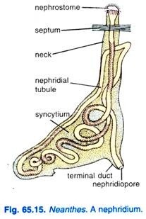
Each nephridium has an oval syncytial mass of protoplasm containing a long, convoluted, ciliated canal, the nephridial tubule. The syncytial protoplasm is differentiated into a body and a neck. The main body is an irregular, oval and compact gland-like mass directed transversely in the segment. It contains highly convoluted and mostly ciliated tubule.
The nephridial tubule passes through the septum into the anterior segment where it opens by a ciliated funnel or nephrostome. Posteriorly the tubule opens by a contractile nephridiopore located ventrally at the base of Para podium near the origin of ventral cirrus. The nephridial tubule is an excavation in the mass of protoplasm. Such an open type of nephridium with a ciliated nephrostome is called a metanephridum.
Physiology of Excretion:
The chief nitrogenous waste in polychaetes is ammonia. Nephridia collect waste from coelomic fluid and blood by diffusion, the cilia of the nephridial tubule cause the liquid waste to pass out of nephridiopores. The waste laden coelomocytes or amoeboid corpuscles are probably removed from the coelomic fluid by the ciliated nephrostomes.
The nephridia of Neanthes Virens also possess the power of osmoregulation.
Its blood and body fluids are isotonic to its surrounding environment over a wide range of salt concentrations, but below a certain limit Neanthes maintains a salt concentration greater than that of the surrounding environment. This maintenance is due to an active intake of salts. Nephridia play an important role in maintaining salt and water balance in the body of Neanthes.
10. Nervous System of Neanthes Virens:
Neanthes Virens has a well developed nervous arrangement. It includes the central, peripheral and visceral nervous system.
(i) Central Nervous System:
Cerebral ganglia or brain is a large bilobed mass, situated in the prostomium above the buccal cavity.
The brain has numerous nerve cells or neurons and nerve fibres and is large in size due to an active life. The middle region of the brain has two small lobes called corpora pedunculata which are association centres and coordinate all the impulses coming to brain.
From the brain arise a pair of stout circumpharyngeal connectives which encircle the pharynx and meet below it in the third segment; close to the brain each circumpharyngeal connective has a ganglion.
Where the circumpharyngeal connectives meet a sub-pharyngeal ganglion formed by the fusion of two pairs of ganglia, it is continuous with a ventral nerve cord made of two cords enclosed in a common sheath. The nerve cord lies below the ventral vessel, it has a double ganglion in each segment beginning from the fourth.
(ii) Peripheral Nervous System:
The peripheral nervous system mainly includes the nerves coming directly from the brain and nerve cord to supply the different regions of the body. The brain gives out four short optic nerves to the eyes, two tentacular nerves to the prostomial tentacles, and two palpal nerves to the palps.
From the small ganglion of circumpharyngeal connectives two pairs of nerves go to the ventral peristomial cirri on each side. From the subpharyngeal ganglion a pair of nerves goes to the first parapodia. Three pairs of nerves arise from each ganglion of the nerve cord, the first goes to the anterior segment and the remaining two pairs innervate the organs of the segment, parapodia and body wall.
(iii) Visceral Nervous System:
The visceral nervous system has two main nerves originating from the sub-pharyngeal ganglion and running parallel to the connectives, each of them has a dorsal and a ventral ganglion; from the dorsal ganglion two pairs of nerves go to the dorsal peristomial cirri, and from the ventral ganglion nerves go to the front part of the alimentary canal.
The nervous system controls and co-ordinates the working of muscles by reflexes, there is a correlation of circular and longitudinal muscles in each segment so that contraction of one layer automatically brings about relaxation of the other. There are nerves between adjacent segments, and excitation of a muscle layer in one segment leads to excitation of the same layer in other segments.
In the nerve cord are five longitudinal giant fibres running along the whole length, they are two large lateral fibres, one large median fibre, and two smaller fibres each of which runs on one side of the median fibre. The giant fibres bring about immediate co-ordination of the entire body because impulses travel rapidly in them from one end of the body to the other, and they cause muscles to contract quickly.
11. Sense Organs of Neanthes Virens:
In Neanthes Virens, the sense organs are specialised and well developed.
On the basis of the function they are of the following types:
(i) Tactile Sense Organs:
We know that the prostomial tentacles, prostomial palpi and peristomial cirri of head are the main tactile sense organs as they are sensitive to touch.
(ii) Chemoreceptors:
It has already been referred to that nuchal organs are a pair of pits on the prostomium, they are lined with ciliated columnar epithelium with some gland cells. These organs are of doubtful nature but some have regarded them as chemoreceptor and olfactory sense organs as being sensitive to taste and smell respectively.
(iii) Photoreceptors:
There are four simple eyes on the dorsal surface of prostomium which are sensitive to light. Each eye has a cup made of pigmented retinal cells produced inwards into clear rods, the opening of the cup is a pupil. In fact, each retinal cell has three distinct parts an outer nucleated part with nerve fibres, a highly pigmented main body and an inner part of transparent cuticular rod.
The external cuticle forms a transparent cornea. Inside the cup is a transparent, gelatinous, refractive lens. The retinal cells are joined to nerve fibres of the optic nerve. The eyes are photoreceptive; they are not related to the formation of image but they help in detecting changes in light intensity.
12. Reproductive System of Neanthes Virens:
Neanthes Virens is dioecious, i.e., sexes are separate. The gonads (testes in male and ovaries in female) are neither permanent nor distinct; they are seasonal and develop only during the breeding season, i.e., in the summer months.
(i) Gonads:
The gonads are formed by the proliferation of the cells of coelomic epithelium and can be seen as groups of masses of germ cells. These groups of germ cells are found particularly on the ventral side in the coelomic space of nearly all segments except a few anterior segments of the body.
However, in male Neanthes dumerilli there is only one pair of testes lying in any one segment between the nineteenth and twenty-fifth segments. But in N. virens and N. diversicolor the testes extend in many segments. In the female the ovaries lie in many segments around blood vessels. Gonads have no gonoducts.
The germ cells from the masses, in male, separate in the coelomic fluid as sperm mother cells, they keep on floating in the coelomic fluid, divide rapidly and undergo maturation show ciliated organs, division to form spermatids which transform into sperms.
A mature sperm has a rod-shaped head and a long vibrating tail. Ova are also formed in the same way in female. An ovum contains yolk granules and is rounded in shape surrounded by a vitelline membrane.
The body of a sexually mature Neanthes is, thus, packed with gametes. Since gonoducts are absent in Neanthes, hence, gametes are discharged out either by the rupture of body wall or by the nephridia. Such nephridia, acting both as excretory and genital ducts, are called mixonephridia (Goodrich, 1945).
(ii) Dorsal Ciliated Organs:
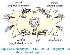
A pair of dorsal ciliated organs is found in each segment situated close to the dorsal longitudinal muscles. Each ciliated organ is a small, ciliated tract of coelomic epithelium and much folded funnel-shaped structure; it opens into the coelom by a wide opening but it has no external opening.
These are believed to be the coelomoducts of other polychaetes and open temporarily to the exterior during breeding season performing the function of gonoducts. However, the true nature of ciliated organs is still to be known.
Neanthes virens and N. dumerilli are extremely variable species, if a number of specimens are examined they show individual variations, these differences are in colour and number of segments. Appearance of orange or reddish colour is the greatest in females during breeding season.
Increase in the number of segments takes place by formation of new segments just anterior to the caudal one. Besides these there may be change in the shape of parapodia, number of their setae, length of the tentacles, and in the number and arrangement of denticles.
Generally a sexually mature Neanthes resembles more or less with non-sexual Neanthes from the morphological point of view.
But in some species certain variations or structural modifications have been recorded in the body of sexually mature Neanthes which are completely lacking in non- mature Neanthes. Such species exhibits two distinct phases in life cycle; a non-sexual or Neanthes phase and sexual or Heteronereis phase. This phenomenon is called epitoky.
(iii) Epitoky and Heteronereis or Heteroneanthes:
Epitoky is a reproductive phenomenon being characteristic of nereids and other annelids like syllids and eunicids. Epitoky is the formation of sexual individual or epitoke which differs from the parent non-sexual individual or atoke.
In fact, these forms differ in such a wide range that sometimes they are considered to be of different genera. Malamgren has recognised that in N. virens, at sexual maturity, the posterior body segments having packed with gametes undergo morphological and anatomical changes.
These changes are induced by hormones secreted into a plexus of blood vessels lying below the brain. Because of such changes the posterior modified segments of the body represent sexual region or epitoke and anterior segments without any change represent non-sexual region or atoke. Thus, the sexually mature Neanthes having these two regions in the body is called Heteroneanthes (= Heteronereis).
However, the Heteronereis is characterised by the following features:
1. It leaves the burrow, comes up at the surface of water and leads an active free swimming life.
2. The eyes become much more prominent, greatly enlarged and highly sensitive to light.
3. The peristomial cirri become longer.
4. The parapodia of epitoke region become enlarged, develop additional foliaceous outgrowths, setae become oar-shaped and highly vascularised.
5. The intestine in the epitoke region becomes compressed due to the development of gonads and finally it becomes functionless.
6. The anal segment (pygidium) develops sensory papillae.
Parapodium of Heteronereis:
The parapodia of epitoke region differ considerably from the parapodia of atoke region.
The parapodia of atoke region resemble the parapodia as described earlier but the parapodia of epitoke region become greatly enlarged, foliaceous, develop additional lobes, dorsal and ventral cirrus are highly enlarged, setae become oar-shaped and arranged in fan-like manner, it becomes highly vascularised.
The original parapodial muscles break down and are digested by leucocytes, then new muscles are formed. Modification of parapodia is an adaptation to a swimming habit and their larger surface serves for more rapid respiration during swimming.
Significance of Heteronereis:
Neanthes Virens is the non-sexual phase and lives in the burrow or creeps at the bottom of the sea, while Heteronereis (= Heteroneanthes) is the sexual phase, swims actively at the surface of sea-water. So, it discharges gametes to far off places in the sea and, thus, helps in the dispersal of species.
13. Swarming of Neanthes Virens:
The fully sexually mature clamworms leave their burrows in groups and come up at the surface of sea-water to swim freely, it is called swarming. This phenomenon probably occurs to facilitate fertilisation. Some species of clamworms are said to perform nuptial dance in which males and females swim rapidly in small circle.
However, swarming occurs at night during a particular lunar phase but the physiological causes and periodicity of swarming are still to be understood. Workers like Clark and Hess (1940) have emphasised that tidal activity and turbidity of sea-water suppress swarming.
14. Fertilisation of Neanthes Virens:
Fertilisation is external and takes place in open sea-water. During swarming, the female Heteronereis produces a substance called fertilisin which attracts the male Heteronereis and it is also responsible for stimulating and shedding of sperms by the males that in turn stimulates female for shedding of ova or eggs (Barnes, 1968). After shedding of gametes the parent worms die.
15. Development of Neanthes Virens:
The development of Neanthes Virens is indirect and can be discussed under the following headings:
Early Embryonic Development:
For making the description easily understandable, it can be discussed as under:
A. Egg:
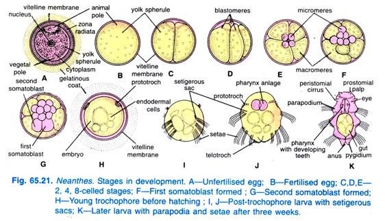
The newly discharged egg of Neanthes is spherical in shape and possesses two membranes; a thin outer membrane which is delicate, followed by a thick radially striated membrane called zona radiata which encloses the ooplasm having yolk spherules, oil droplets and female nucleus. An egg is surrounded in a thick, transparent, gelatinous coat.
However, immediately after fertilisation, several changes occur in an egg; the zona radiata disappears, yolk spherules aggregate at the lower end of the egg leaving a clear zone in the upper end. The lower end, thus, becomes to be known as vegetal pole and the upper end as animal pole; the egg is, thus, telolecithal. Two small polar bodies are extruded towards the animal pole and the zygote undergoes cleavage.
B. Cleavage and Formation of Germ Layers:
The zygote undergoes cleavage in which the first two divisions are equal producing four cells which are equal and lie in the same plane, these cells are A, B, C and D. Each cell gives rise to one of the quadrants of the embryo. D is larger than the others and forms the dorsal side of the embryo, B is ventral, and A and C are lateral.
The succeeding cleavages are unequal and at right angles to the first two divisions, they form three quartets of micromeres which are divided off from the fourth quartet of macromeres, the micromeres lie towards the animal pole and the macromeres, at the vegetal pole.
The micromeres are not directly over the macromeres, but one quartet is displaced to the right, the next is displaced to the left, and the next to the right again.
This pattern of division is called spiral cleavage in which any one cell lies between two blastomeres or below it, and at first the cleavage planes are oblique to the polar axis (axis between animal and vegetal poles). Later the successive cleavage planes are at right angles.
Spiral cleavage is determinate, if at the 4-cell stage the blastomeres are separated, then each will form only one quarter of the embryo, that is the fate of blastomeres is predetermined, this is known as determinate cleavage.
The fate of blastomeres is fixed and each will give rise to a particular tissue only. The cells of the first three quarters give rise to the ectoderm of the larva and the adult.
At the next cleavage a fourth quartet is separated from the macromeres, one of the cells of the fourth quartet known as a somatoblast produces the entire mesoderm, while the remaining three cells reinforce the macromeres and all of them form the endoderm. The cells eventually form a gastrula, then the three germinal layers are formed.
Structure of Trochophore and its Metamorphosis:
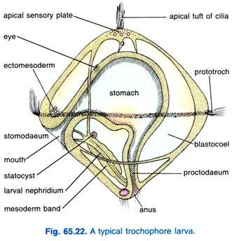
The gastrula develops into a trochosphere or trochophore larva. The trochosphere is not only characteristic of Polychaeta, but it also occurs is Mollusca, Archiannelida, and polyclad Turbellaria. The trochosphere (Fig. 65.22) is rounded and transparent, it has a thin external ectodermal epithelium which is thickened at the two ends and along an equatorial ring.
There is a curved gut with a mouth, ectodermal oesophagus or stomodaeum, an stomach, and an hindgut opening by an anus. It feeds on micro-organisms. On the thickened parts of the ectoderm is an anterior ciliated apical organ with an apical ganglion below which is an eye spot, at the posterior end are some large cilia and on the equatorial ring is a pre- oral ciliated band or prototroch.
Between the ectoderm and gut is a large cavity, the blastocoel having mesencyme cells, larval mesoderm and a pair of larval nephridia, each made of two hollow cells, one of which contains a flame of cilia, there is an otocyst near the nephridia.
The trochosphere is pelagic, it drifts about in the sea swimming by its prototroch ; organs of the adult begin to form. The apical organ forms the prostomium with brain, tentacles and eyes. The part immediately behind forms the peristomium. The larva grows from the anal end as an elongated cylinder which forms segments of the body by metameric segmentation.
The larval nephridia are replaced by permanent ones, larval setae are dropped, tentacles, palps and parapodia are formed.
The advanced larva consists of the adult head and body segments separated by the body of the larval trochosphere, this larval region shrivels up, and the head and body segments are drawn together and joined to metamorphose the larva into a young worm. The young worm, thus, resulted, settles at the bottom of the sea and starts forming its burrow and gradually attains adulthood.
However, it is interesting to note that a typical free-swimming trochophore, as described above, occurs only in some polychaetes. In Neanthes Virens, this stage is passed inside the egg membrane and the larva which hatches out of the egg is advanced trochophore larva referred to as nectochate which undergoes metamorphosis to become a young worm in the same way as described above.


