In this article we will discuss about the reproductive system of cockroach.
Male Reproductive Organs of Cockroach:
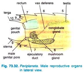
The male reproductive system of cockroach consists of a pair of testes, vasa deferentia, an ejaculatory duct, utricular gland, phallic gland and the external genitalia.
(i) Testes:
There is a pair of three-lobed testes lying dorsolaterally in the 4th and 5th abdominal segments, being embedded in the fat body. The testes are well developed and elaborate structures in young cockroach and they are full of sperms. The testes become non-functional and reduced in old adults but some sperms may still be found in them.
(ii) Vasa Deferentia:
From each testis arises a thin thread-like, white vasa deferens. Both the vasa deferentia pass backwards almost to the posterior end of abdomen and then bend forwards to meet in the middle and open into an ejaculatory duct.
(iii) Ejaculatory Duct:
The ejaculatory duct is an elongated wide median duct which runs backwards in the abdomen and opens out by male gonopore situated ventral to the anus.
(iv) Utricular or Mushroom-shaped Gland:
It is a large accessory reproductive gland, whitish in colour and situated at the junction of vasa deferentia with the ejaculatory duct. It has a mass of glandular tubules of three kinds, the peripheral long tubules or utriculi majores, the central tubules are small short tubules or utriculi breviores and behind the short central tubules are some short but more bulbous tubules forming the seminal vesicles filled with sperms.
(v) Phallic or Conglobate Gland:
It is a long and club-shaped accessory gland. Its anterior broader end lies in the 6th segment slightly to the right of the nerve cord. It narrows posteriorly into a tubular structure and finally tapers to open by a separate aperture located close to the male gonopore at the hind end of the body.
(vi) External Genitalia:
Some chitinous asymmetrical structures are found surrounding the male gonopore at the end of the abdomen. These are three phallomeres or male gonapophyses which constitute the external genitalia.
Right Phallomere:
It is mid-dorsal in position. It has two chitinous but membranous horizontal opposing plates and a broad serrate lobe with a saw- toothed edge and two large teeth, and at its posterior side it has a sickle-shaped hook.
Left Phallomere:
It has a broad base from which several structures arise, on the extreme left is a long slender arm with a curved hook called titillator, next to the titillator is a shorter and broader arm ending in a black hammer-like head called pseudopenis.
Close to the pseudopenis are three small soft lobes, one of which bears a hook and is called an asperate lobe. The duct of the phallic gland traverses the left phallomere and opens between the asperate lobe and pseudopenis.
Ventral Phallomere:
It is very simple in structure and lies partly below the right phallomere. It has a large brown plate and bears the male gonopore.
Spermatophore:
The sperms produced from testes, while the cockroach is still young, are brought by the vasa deferentia into the seminal vesicles for storage. The sperms in the seminal vesicles are glued together in the form of bundles called spermatophores. Actually, the spermatophores are discharged by the male during copulation.
A spermatophore is pear-shaped about 13 mm in diameter and its wall has three layers.
Its innermost layer is first formed by the milky secretion secreted from the long peripheral tubules of the utricular gland. This layer then receives bundled sperms from seminal vesicle and a liquid from the short tubules of the utricular gland. Then this inseminated layer passes down the ejaculatory duct and it receives the second layer from the cells of ejaculatory duct.
During mating, the two layered spermatophore, thus, formed is attached to the spermathecal aperture of the female and then the secretion of phallic gland is poured over it which hardens to form the third and outermost layer of the spermatophore (Fig. 73.34).
Female Reproductive Organs of Cockroach:
The female reproductive system of cockroach (Fig. 73.35) consists of a pair of ovaries, vagina, genital pouch, collaterial glands, spermathecae and the external genitalia.
(i) Ovaries:
There are two large, light yellow- coloured ovaries lying laterally in the segment 4th, 5th, 6th, embedded in the fat body. Each ovary is formed of a group of eight ovarian tubules or ovarioles containing a chain of developing ova. An ovariole is made up of an epithelial layer resting on a basement membrane and enclosed externally in a connective tissue coat.
However, an ovariole from in front to backwards consists of the following zones:
(i) Suspensory filament, it is thin, thread-like continuation of the connective tissue layer and provides attachment of the ovariole to the dorsal body wall and, thus, it serves to suspend the ovariole in the haemocoel.
(ii) Zone of germarium, it follows the terminal filamentous zone and consists of germ cells or oogonia and mature into oocytes and pushed downwards.
(iii) Vitellarium, this zone receives the oocytes from the zone of germarium one by one and constitutes the largest part of the ovariole, the oocytes become enclosed in a follicle of epithelium and increase progressively in size towards the posterior end which gives it beaded appearance.
(iv) Egg chamber, the vitellarium opens posteriorly into a small, thick, oval egg chamber which contains a single large mature ovum at a time.
(v) Stalk or pedicel, the egg chamber continues posteriorly into thin-walled, hollow stalk which opens into the lateral oviduct.
Oviducts:
The stalk of all eight ovarioles on one side join to form an oviduct which is lateral, small and with muscular wall.
(ii) Vagina:
Both the lateral oviducts unite to form a broad median common oviduct called vagina. The vagina opens by the female gonopore into the genital chamber.
(iii) Genital Pouch:
It is a large boat-shaped structure whose floor is formed by the 7th sternite, roof and sides are formed by the 8th and 9th sternites. The genital pouch can be divided into a genital chamber into which vagina opens and an oothecal chamber where oothecae are formed. The genital chamber also receives the accessory reproductive glands.
The female gonopore is an aperture in the 8th sternum, which lies inside the genital chamber inflected above the 7th sternite. The 7th sternite is also produced backwards into two large oval gynovalvular plates or apical lobes. The genital pouch is also referred to as gynatrium.
(iv) Collaterial Glands:
There is a pair of white much branched collaterial glands, the left is much larger than the right. Both these glands continue as collaterial ducts which join to form a common duct which opens into the dorsal side of the genital chamber. These are the accessory reproductive glands.
(v) Spermathecae:
These are a pair of club-shaped, unequal-sized, one spermathecae being larger than the other, structures. Both the spermathecae unite to form a short common duct which opens into the genital chamber on a small spermathecal papilla. Some workers claim that there is a single spermatheca and it has a lateral caecum.
(vi) External Genitalia of Female:
These lie concealed inside the gynatrium. They consist of an ovipositor formed by two gonapophyses. The ovipositor lies above and behind the gonopore, it is short and has three pairs of elongated processes, a pair of long thick arms lying dorsally and enclosing two pairs of slender tapering arms.
These two pairs of arms arise from a common base and they constitute the posterior gonapophyses, they belong to the 9th abdominal segment and are joined to the 9th tergum.
The third pair of arms of the ovipositor is large, they converge and meet posteriorly lying below the posterior gonapophyses and constitute the anterior gonapophyses. These belong to the 8th abdominal segment and are attached to the outer margins of 8th tergum. The ovipositor is used only to conduct fertilised eggs to the oothecal chamber.


