In this article we will discuss about the functions of male and female reproductive organs, explained with the help of suitable diagrams.
Male Reproductive Organs:
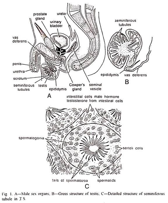
Sperms are produced in the testes. The testes are two oval bodies and are suspended in a sac hanging from the lower wall of the abdomen, the scrotum. Each testis is composed of coiled anastomosing seminiferous tubules lined with epithelial cells that produce sperm cells; also interstitial cells of Leydig around the tubules produce the male sex hormone, testosterone, which promotes the development of the accessory glands and controls male secondary sex characteristics.
As sperms are released into the interior of the tubules they are carried by ciliary action to the epididymis which lies on the outride of and partially encircling the testis. Testis and epididymis together constitute testicle. In the epididymis the sperms are stored so that they become motile.
Epididymis connects with the vas deferens. Vas deferens is a muscular tube that leaves the scrotun by the inguinal canal and empties into the urethra, the duct that leads from the bladder. The terminal portion of each vas deferens enlarges to form an ejaculatory duct, capable of contraction and expulsion of the sperms which are stored there.
A glandular seminal vesicle empties into each ejaculatory duct before it connects to the urethra. The seminal vesicles secrete a viscid fluid which is expelled along with sperms. The mixture of this fluid and the sperms is known as semen (Fig. 1).
The urethra is surrounded by a prostate gland at the point where the ejaculatory ducts enter. This gland has numerous small ducts emptying into the urethra. Its secretions are thin milky in nature containing citiric acid, calcium, phosphate fibrinogenase, fibrinolysin, spermin etc. and contributes 15-30% of the total-volume of the semen.
Another pair of glands, the Cowper’s glands, are also attached to the urethra below the prostate gland. Their secretions are also alkaline and serve as lubricant for the semen. The secretions of the prostate and Cowper’s glands suspend the sperms, motile them, nourish them and neutralize the normally acid environment of the urethra and of the female reproductive tract to a pH more suitable for sperm survival.
Urethra communicates with the exterior of the body through a muscular structure, the penis. The penis consists of columns of spongy tissue, the corpora cavernosa, surrounding the urethra, and a layer of skin on the outside. The tip of the penis enlarges slightly to form the glans, which is normally covered by a fold of skin, the prepuce. The function of penis is to deposit the semen in the genital tract of the female.
Erection of Penis:
It is associated with sexual stimulation. It is caused by dilation of the blood vessels carrying blood to the spongy tissues, resulting in the collection of blood within these spaces. As the tissues become distended they compress the veins and so inhibit the flow of blood out of the tissues. With continued stimulation the penis and the underlying bulb become hard and enlarged.
Ejaculation:
Continued stimulation of the penis leads to contraction of the muscles present in the scrotun, raising the testes close to the body, epididymis and vas deferens. These contractions move the semen into urethra. Finally the muscles surrounding the bulb are stimulated. They contract and propel the semen out through the urethra and produce some of the sensations associated with orgasm.
Semen:
It is a fluid which is ejaculated at the time of insemination. It contains sperm cells and secretions of seminal vesicles, prostate gland and Cowper’s gland and also of urethral glands. The amount of semen discharged per ejaculation varies from 2.5 to 3.5 ml.
Spermatozoon:
The spermatozoon of man consists of two parts, head and tail. The tail is divided into neck, mid-piece, principle-piece and end-piece. It is about 0.05 mm in length. It is motile in nature and enzymes that are responsible for its motility are located in the mid-piece (Fig. 2).
The head of spermatozoan is a spoon-shaped structure which is bounded externally by plasma membrane. At the anterior end it has a cup-like structure called acrosome made up of Golgi apparatus. It contains hydrolyzing enzymes and plays an important role in the penetration of sperm in the ovum. Head in its interior contains a well condensed nucleus and a very little cytoplasm. Head is followed by a short neck.
Neck consists of a pair of centrioles, a proximal centriole and a distal centriole. The two centrioles lie at right angles to one another. The proximal centriole has no active function but is a potential activist within the egg during the first cleavage division of the fertilized egg. The distal centriole serves as basal body for the tail.
Neck is followed by mid-piece which is composed exclusively of mitochondria which aggregate about its basal end, forming a continuous spiral. This mitochondrial apparatus provides a ready energy source (ATP) to the sperm tail for its motility, the mid-piece is followed by the principal-piece which ultimately ends in an end-piece. In principal-piece and end-piece the fibre-system is reduced to the axial complex of two central fibres surrounded by the ring of nine peripheral fibres.
Numbers:
About 300 to 400 millions of sperm cells are present in the semen of each ejaculate of a normal young adult male but of those only one can fertilize each egg cell. Males that produce less than 35 millions of sperms per millilitre of semen are generally sterile.
Female Reproductive Organs:
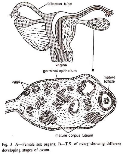
The female reproductive organs include a pair of ovaries, a pair of oviducts (Fallopian tubes), the uterus and the vagina (Fig. 3).
The human ovaries are two small almond-like flattened bodies lying on the sides of vertebral column behind the kidneys in the pelvic cavity. Each ovary is attached to the dorsal abdominal wall through mesovarium and ovarian ligament. Each ovary is principally composed of stroma of fibrous connective tissue and is lined externally by a germinal epithelium which proliferates thousands of primordial follicles during the embryonic life of an individual.
Each ovary is roughly differentiated into an outer cortex and an inner medidla. In mature ovary the cortex contains follicles and corpora lutea in various stages. The medulla consists only the large blood vessels of the organ. One cell of the mass of epithelial cells gives rise to an immature ovum or oocyte; the remaining cells form a layer surrounding the ovum or oocyte as sac or follicle called follicular epithelium or granulosa.
Immature ovum or oocyte and surrounding follicular epithelium or granulosa constitute the primordial follicle. The stroma of the ovary surrounding the follicular epithelium or granulosa becomes organized into connective tissue layers, the theca externa and the theca interna.
Puberty:
In the young girl the Graffian follicles do not reach maturity. They begin to ripen only when the puberty commences in the girl (10-15 years).
Puberty is the period of onset of sexual maturity or sex organ function and beginning of youth. During this period, morphological, psychological, physiological and endocrinological changes take place in the individual.
The accessory sex organs, uterus, vagina, and mamma undergo a marked increase in growth. Secondary sexual characters like pubic and axillary hair, the peculiar development of the skeleton and deposition of fat on the hips which causes the body to assume a more famine contour.
All changes undergone by a young girl during puberty are due to pituitary gonadotropins which stimulate the ovaries. Ovaries on stimulation secrete some specific hormones especially the progesterone and oestrogen which regulate the development and maintenance of primary as well as accessory sex characters of the female individual.


