In this essay we will discuss about Cockroach. After reading this essay you will learn about: 1. Habitat and Habits of Cockroach 2. Morphology of Cockroach 3. Anatomy 4. Endoskeleton 5. Digestive System 6. Digestive Glands 7. Heart 8. Respiratory System 9. Mechanism of Respiration 10. Excretory System 11. Nervous System 12. Endocrine System 13. Sense Organs 14. Compound Eyes and Other Details.
Essay Contents:
- Essay on the Habitat and Habits of Cockroach
- Essay on the Morphology of Cockroach
- Essay on the Anatomy of Cockroach
- Essay on the Endoskeleton of Cockroach
- Essay on the Digestive System of Cockroach
- Essay on the Digestive Glands of Cockroach
- Essay on the Heart of Cockroach
- Essay on the Respiratory System of Cockroach
- Essay on the Mechanism of Respiration of Cockroach
- Essay on the Excretory System of Cockroach
- Essay on the Nervous System of Cockroach
- Essay on the Endocrine System of Cockroach
- Essay on the Sense Organs of Cockroach
- Essay on the Compound Eyes of Cockroach
- Essay on the Reproductive System of Cockroach
- Essay on the Interaction of Cockroach with Mankind
Essay # 1. Habitat and Habits of Cockroach:
Cockroaches are found in warm, dark and damp places. They commonly inhabit kitchens, restaurants, store houses, godowns, railway wagons, ships, etc. They are numerous in underground drains.
They are nocturnal insects preferring darkness and become active during night, but remain hidden under some objects and take rest during day time.
Cockroaches are omnivorous in diet, feeding on almost all kinds of food matter including human food, paper, leather, cloth and even dead bodies of their fellows (show cannabilism). They prefer starch containing food. Cockroaches are cursorial insects, viz., run very fast. They fly very rarely as they are not good fliers.
Cockroaches are unisexual, viz., male and female sexes are found in different individuals. They show sexual dimorphism, viz., one sex can be distinguished from the other externally. Cockroaches are oviparous, viz., they lay eggs. Parental care is seen in these insects.
The young cockroaches, called the nymphs, resemble the adults in most of the characters, but are smaller in size, pale in colour, devoid of wings. Nymphs undergo moulting or ecdysis, in which the casting of older skin takes place. The nymphs gradually change into adults.
Lizards, birds, moles and even toads are the chief enemies of the cockroaches.
Essay # 2. Morphology of Cockroach:
Shape, Size and Colour:
The body is dorsoventrally flattened, elongated and bilaterally symmetrical. The adult cockroach is about 34-53 mm long with wings that extend beyond the tip of the abdomen in males.
Exoskeleton:
The body of cockroach is externally covered by hard brown chitinous plates, the clerites, which constitute the exoskeleton. The sclerites are joined with one another by thin flexible, soft articular or arthrodial membranes.
Body Divisions:
The body of cockroach is distinctly divided into three regions, viz., head, thorax and abdomen.
I. Head:
It is the anterior most region of the body, which lies at right angle to the long axis. It is formed by the fusion of six segments. The proximal semi-circular broader end of the head is directed upwards, while the distal narrow, mouth bearing end is directed downwards.
This type of head is called hypognathous. The neck allows the movement of the head in all possible directions. Head is covered by sclerites and bears sense organs, mouth parts and mouth.
Sclerites:
In adult all sclerites of the head are fused to form a head capsule. The top of the head capsule is known as vertex. The latter is divided by ‘λ’ shaped epicranial suture into two epicranial plates. A triangular plate, the frons is found in front of the epicranial plates.
The frons is followed by a rectangular plate, the clypeus. A frontoclypeal suture is present in between the frons and clypeus. A movable chitinous flap, the labrum hangs from the lower edge of the clypeus. A labroclypeal suture is found in between the clypeus and labrum. Two cheek sclerites, the genae, lie vertically just below the eyes at the lateral side.
Sense organs:
The sense organs present on the head are compound eyes, antennae and fenestrae or ocellar spots.
(a) Compound eyes:
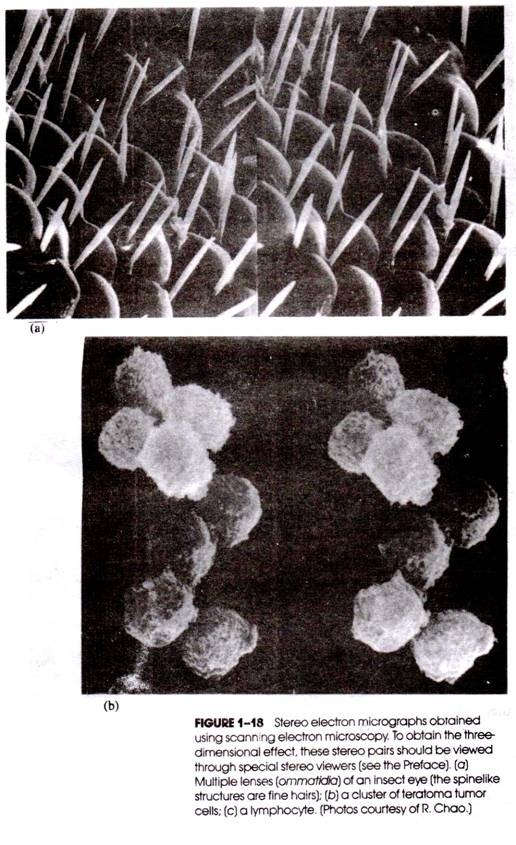
Two large black coloured, kidney shaped, dorsolaterally placed compound eyes lie on the top of the head. They are formed by a large number of visual elements, the ommatidia (sing ommatidium). The latter are externally represented by the same number of hexagonal areas. The compound eyes are the organs of sight.
(b) Antennae:
A pair of filifrom (like a thread) antennae arise from antennal sockets lying in front of eyes. The antenna is made up of many segments which are called podomeres. The antennae can be moved in all directions. They possess sensory bristles, which are tactile in nature. With the help of antennae, the cockroaches can detect the presence of food and the object in front. Antennae also respond to smell.
(c) Ocellae or Fenestrae:
A small rounded pale- coloured area, the ocellus or fenestra is situated just towards the inner and upper side of each antennal socket. They represent the undeveloped simple eyes. They are sensitive to light.
Mouth parts:
The mouth lies at the lower part of the head, and the appendages associated with the mouth are called mouth parts. They are used in searching and taking the food matter. The biting and chewing type of mouth parts of cockroach consist of a labrum, two mandibles, two maxillae, a labium and a hypo pharynx.
(i) Labrum:
Labrum is also called upper tip. It is a broad, chitinous flap, which hangs from the distal end of the clypeus. A thin plate, the epipharynx, is fused to the inner surface of the labrum. The labrum holds the food particles during the feeding.
(ii) Mandibles:
This part of a into mandible is divisible into grinding region and incising region. These are two stout, structures which lie below the genae. Inner margin of each mandible bears teeth, while on its upper inner edge, a soft lobe, the prostheca, is present.
When both the mandibles work simultaneously in a horizontal plane, the food matter is cu and masticated into fine and smaller pieces. Mandibles move with the help of adductor and abductor muscles.
(iii) Maxillae (First Maxillae):
They lie beneath the mandibles one on either side of the head capsule.
Each maxilla consists of three parts:
(a) Protopodite:
It is a basal portion and made up of two parts: the proximal cardo and the distal stipes. Both the parts are bent at an angle to each other,
(b) Endopodite:
It arises from the inner side of the stipes and consists of two parts: outer broad, hood-like galea and an inner, hard plate-like lacinia. The latter tapers into two sharp claw like projections. Lacinia also bears numerous strong sensory bristles at its inner surface,
(c) Exopodite:
It consists of a small basal sclerite, the palpifer attached to the outer surface of the stipes and a five jointed maxillary palp with sensory bristles. With the help of lacinia, it holds the food and gives it to the mandibles for mastication. Maxillae are also used for cleaning the antennae and the first pair of legs. The maxillary palps respond to touch, taste and smell.
(iv) Labium (Second Maxillae):
Labium is also called lower lip. It is a single structure which represents the fused second pair of maxillae, lying behind the mouth and forming a sort of lower lip.
It comprises the following parts:
(a) Protopodite:
It consists of a proximal large part, the submentum, the middle small mentum and the distal pre-mentum. The sub-mentum and mentum are together called as post-mentum, which probably represents the fused cardos, while pre-mentum is perhaps the fused portion of two stipes,
(b) En- dopodite:
Partially fused endopodites are represented by the ligula whose each half consists of an inner glossa and an outer paraglossa, which correspond to the lacinia and galea of the first pair of maxilla, (c) Exopodite. It consists of two parts.
A small sclerite, the palpiger, arises from the pre-mentum on each side and a three jointed labial palp, bearing sensory bristles attached to the palpiger. It corresponds to the maxillary palp of the first maxilla. The labium does not take active part in feeding. However, glossae and paraglossae pi event the loss of food particles from mandibular action. The labial palps respond to taste and smell.
(v) Hypopharynx:
It is a small conical fleshy structure, hanging in between the two maxillae in front of the labium and acts like tongue. An efferent salivary duct carrying the saliva from the salivary glands opens near the base of the hypopharynx. Hypopharynx directs the saliva towards the food in the preoral cavity.
Mouth:
It is a narrow opening that lies at the base of the pre oral cavity. It is bounded by the mouth parts. It leads into the pharynx.
II. Thorax:
It consists of three segments. The anterior pro-thorax, middle mesothorax and posterior metathorax.
(i) Thoracic Sclerites:
Each thoracic segment is enclosed by four chitinous skeletal sclerites, a dorsal tergum, a ventral sternum and two lateral pleura (sing, pleuron). The tergum of the pro-thorax is also called pro-notum, which is the largest sclerite and projects forwards to cover the neck.
(ii) Thoracic Appendages:
The thorax bears three pairs of legs and two pairs of wings.
(a) Legs:
Each thoracic leg segment bears a pair of walking legs. According to their position, they are named as pro-legs, meso-legs and meta-legs. They arise in between the sterna and pleura. All the three pairs of jointed legs are similar in structure. Each leg is composed of five podomeres. Coxa.
It is a small, hard podomere, which articulates with the thoracic segment in between the pleuron and the sternum. Trochanter. It is a small, hard podomere which lies next to the coxa. Trochanter articulates movably with the coxa, but is fused with the next podomere, the femur.
Femur it is a long and the stoutest part of the leg, bearing sensory bristles. Tibia it is a thin, slender and the longest part of the leg, which also possesses bristles. Tarsus it is a terminal part of the leg and composed of five tarsomeres.
The terminal tarsomere is known as pretarsus, which bears two sharp curved claws and a soft hairy pad, the pulvillus, in between the two claws. Each tarsomere possesses a soft adhesive pad, the plantula on its lower side. The claws and pads help the cockroach in grasping the substratum firmly.
(b) Wings:
Two pairs of wings are found in cockroach. The first pair is mesothoracic and attached with the tergum of the mesothorax. The second pair is metathoracic connected with tergum of metathorax. In fact, the wings are the membranous outgrowths of the body wall, supported by a network of slender and branched tubules, called nervures or veins.
These tubules communicate with haemocoel. Thus haemocoelomic fluid circulates in these tubules. An air tube or trachea and a fine nerve also present in each nervure. The movement of the wings is controlled by a special set of muscles.
Mesothoracic Wings (Forewings):
They are thick, leathery, opaque and dark coloured structures, which are somewhat narrow at the distal end. They are larger than the second pair of wings. They are not used for flight, but cover and protect the metathoracic wings. Mesothoracic wings are attached with the anterolateral margin of the mesothoracic tergum. They are also called wing-covers or tegmina or elytra.
Metathoracic Wings (Hind Wings):
They are delicate, thin, transparent and membranous structures with broad terminal end. They are used for flight, but in the resting position, lie folded below the tegmina. Metathoracic wings are attached with the anterolateral margin of the metathoracic tergum.
(iii) Thoracic Spiracles:
There are present two pairs of thoracic spiracles. The first pair of spiracles is known as mesothoracic, lying in front of the mesothorax between the bases of first and second pairs of legs. These are the largest spiracles. The second pair of spiracles is called metathoracic, lying in front of the metathorax, between the bases of second and third pairs of legs. Spiracles are meant for intake of fresh air and release of foul air.
III. Abdomen
It is the largest part of the body narrows posteriorly. The abdomen in both male and female consists of 10 segments.
(i) Abdominal Sclerites:
A typical abdominal segment is enclosed by four sclerites. A dorsal tergum, one ventral sternum and two lateral pleura are found in a typical thoracic segment. There are present ten terga. The 7th tergum covers the 8th tergum in male and 8th and 9th terga in the female cockroach. The 10th tergum is large and notched in the middle and projects backwards beyond the body.
The abdomen bears 9th sterna, while the 10th one is absent. In male all the 9 sterna are seen, but in female cockroach only 7 sterna are visible, the 7th one conceals the 8th and 9th sterna. The abdomen in both male and female cockroaches consists of 10 segments.
In females, the 7th sternum is boat shaped and together with the 8th and 9th sterna forms a gential pouch (brood pouch) which is divisible into a genital chamber in-front and vestibulum (oothecal chamber) behind.
Genital chamber contains female gential pore, spermathecal pores and collaterial glands. In males, genital pouch lies at the hind end of abdomen. It contains dorsal anus ventral male gential pore and gonapophyses (phallomeres). The abdomen of female cockroach is broader than the male.
(ii) Abdominal Appendages:
The posterior end of the abdomen bears the following appendages:
(a) Anal cerci:
These are paired, jointed outgrowths, which arise from the 10th tergum. The anal cerci are long and thick structures found in both male and female cockroach and are sensitive to touch. They are also sensitive to sound and other vibrations,
(b) Anal styles:
These are also paired but thin, small un-jointed outgrowths, which project backwardly from the sides of the 9th sternum of the male cockroach only. They are also sensitive to touch,
(c) Gonapophyses:
In both the sexes the genital aperture is surrounded by some sclerites, known as gonapophyses. In male, they arise from the 9th segment and form the external genitalia or external genital organs to help the insect in copulation. In the female, gonapophyses belong to the 8th and 9th abdominal segments to form an ovipositor. The latter is used to guide the fertilized eggs towards the oothecal chamber for ootheca formation.
(iii) Stink glands:
A pair of stink glands is present between the fifth and sixth abdominal terga. These glands produce a secretion that gives a characteristic stinky (foul) smell.
(iv) Apertures:
The abdomen bears the following apertures:
(a) Anus:
The waste matter is expelled out from the posterior most part of the alimentary canal, the anus,
(b) Genital Aperture:
In the male it lies below anus. Female genital aperture opens into the genital chamber. In both the sexes genital aperture is surrounded by gonapophyses.
(c) Abdominal Spiracles:
There are present eight pairs of abdominal spiracles. The first pair of abdominal spiracles is dorsal in position and lies on the lateral margins of the first abdominal tergum.
The remaining abdodminal spiracles are situated on the sides of their corresponding segments on the pleura between the terga and sterna. The wall of the spiracles is provided with fine bristles to check the entry of dust particles while oxygen enters the respiratory tubes, the tracheae, for respiration.
Essay # 3. Anatomy of Cockroach:
Body Wall (Fig. 7A.20):
Body wall of cockroach consists of the following three layers.
(i) Cuticle:
It forms the outermost layer of the body wall secreted by the epidermis. It is made up of chitin which becomes hard to form the exoskeleton.
The cuticle consists of three sub-layers:
(a) Epicuticle:
It is outer, thin and waxy layer which is usually impermeable to water. Chitin is absent in epicuticle.
(b) Exocuticle:
It is the middle laminated, tough, chitinous pigmented thinner layer,
(c) Endocuticle:
It is a thick innermost layer formed of soft laminated chitin. The cuticle forms some immovable spines which are tactile in function.
(ii) Epidermis (= Hypodermis):
It lies below the cuticle and consists of single layered columnar cells. It secretes the cuticle. Certain cells of the epidermis are modified into trichogen cells which secrete movable bristles. Tormogen cells secrete membranes around the bristles.
Dermal gland cells secrete waxy substance which is spread over the cuticle and gives obnoxious smell. Oenocytes produce materials for the formation of the epicuticle. Oenocytes also influence moulting (ecdysis).
Numerous fine pore canals which are spirally coiled tubes traverse vertically through the endocuticle and exocuticle of the body wall of cockroach. These are the ducts of the glandular cells of underlying epidermis. They are supposed to influence flexibility and permeability of cuticle.
(iii) Basement membrane:
It lies beneath the epidermis. It is composed of flattened cells.
Essay # 4. Endoskeleton of Cockroach:
At some places, certain processes (projections) of exoskeleton extend into the body and form endoskeleton elements. These provide attachment to the muscles and hence called apodemes.
(i) A tent-like plate called tentorium, forms the endoskeleton of head,
(ii) In the thorax, separate processes of stemites of the three segments form endoskeleton.
(iii) Abdomen of cockroach does not have endoskeleton elements.
Body Cavity:
True coelom is found during embryonic development. In adults, original coelom is present around the gonads. The body cavity is filled with blood and, therefore, called haemocoel. The haemocoel is divided by two horizontal perforated partitions or diaphragms into three spaces, the pericardial, perivisceral and perineural sinuses.
Fat Body (= Corpora Adiposa):
Fat body fills most of the perivisceral sinus around the gut. Fat body consists of numerous solid lobules. It is bound by a fine membranous covering of connective tissue. Each lobule contains several types of cells. Four cell types are of special significance.
(i) Trophocytes:
They contain reserve food in the form of fats, glycogen and protein,
(ii) Mycetocytes:
These cells contain symbiotic bacteria which help in synthesis of some amino acids and vitamins and of glycogen from glucose,
(iii) Oenocytes:
They are probably concerned with some intermediary metabolism at time of ecdysis.
(iv) Urate cells:
They absorb nitrogenous waste products from the haemolymph and synthesize uric acid from these for storage.
Essay # 5. Digestive System of Cockroach:
The cockroach has well developed digestive system comprising the alimentary canal and associated digestive glands.
Alimentary Canal:
It consists of mainly three parts; fore-gut or stomodaeum, midgut or mesenteron and the hind-gut or proctodaeum. The fore and hind-guts are formed by ectoderm and are lined by the cuticle internally, while the mid-gut development is from endoderm of the embryo and not lined by the cuticle.
I. Stomodaeum or fore-gut:
It comprises the pre-oral cavity, mouth, pharynx, oesophagus, crop and gizzard.
(i) Pre-oral cavity:
It is a space lying in front of the mouth to receive the food, hence named as pre-oral cavity. The cavity is bounded anteriorly by the labrum, posteriorly by the labium, laterally by the mandibles and maxillae. Inside the cavity, a tongue like structure, the hypo pharynx projects from the posterior wall of the preoral cavity. The hypo pharynx bears at its base an opening of efferent salivary duct.
(ii) Mouth:
A narrow opening, the mouth lies at the base (bottom) of pre-oral cavity which is bounded by the mouth parts. Mouth leads to the pharynx.
(iii) Pharynx:
It is a short tube-like structure which lies in the head region and leads into the oesophagus.
(iv) Oesophagus:
It is a tubular thin walled structure and succeeds anterior part of the thorax. It continues into the crop.
(v) Crop:
The oesophagus dilates to form a large thin walled pear shaped crop. The crop is very large part of the alimentary canal of the cockroach where food is stored. It opens into the gizzard.
(vi) Gizzard (= Proventriculus):
It is a thick walled somewhat rounded structure whose walls are muscular and greatly folded. It has six teeth, used for grinding the food. Its wall consists of strong circular muscles which are needed by the gizzard for grinding.
II. Mesenteron or mid-gut:
It is a thin walled somewhat coiled tube with almost uniform thickness. Main digestion and absorption of food is carried out here. A peritrophic membrane is formed around the food in the mid gut. This membrane is permeable to digestive enzymes and digested foods.
From the junction of mid-gut and gizzard arise six to eight finger-like blind structures called the hepatic caecae or gastric caecae. The junction of mid-gut and hind-gut is marked by the presence of 100—150 yellow fine thread-like structures, the Malpighian tubules. The tubules are excretory in function.
III. Proctodaeum or Hind-gut:
It comprises the ileum, colon, rectum and anus. It is ectodermal in origin. Hind gut is broader than mid gut.
(i) Ileum:
The mid-gut continues into the ileum. It is short and relatively narrower. Anteriorly it bears thread like Malpighian tubules. The ileum passes undigested food into the colon.
(ii) Colon:
Actually it is a dilation of ileum and is the longest, relatively thicker and coiled part of hind gut. It leads into the rectum.
(iii) Rectum:
It is the terminal short thick walled structure. Its internal lining is thrown to form six thick longitudinal folds known as rectal papillae. The latter help in the absorption of water.
(iv) Anus:
It is a slit like opening of the alimentary canal lying at the hind end of the abdomen below the 10th tergum.
Essay # 6. Digestive Glands of Cockroach:
They are as follows:
(i) Salivary glands:
There are paired salivary glands lying one on each side of the oesophagus and crop. Each gland has two glandular portions and a salivary reservior. The secretion secreted by the glandular portions is called saliva which is stored in the reservoirs and is carried to the preoral cavity through efferent salivary duct. The saliva contains amylase, chitinase and cellulase enzymes.
(ii) Hepatic caecae:
There are 6 to 8 tubular structures arising from the anterior end of the mid-gut. Hepatic caecae are lined by the glandular cells which secrete digestive secretion containing amylolytic, proteolytic and lipolytic types of enzymes.
(iii) Mid-gut lining:
The mid-gut is lined by glandular epithelial cells which secrete a digestive secretion containing proteolytic, amylolytic and lipolytic enzymes.
Food:
Cockroach is omnivorous. Its food consists of almost all types of organic matters, i.e., paper, bread, cloth, vegetables, meat, etc. It also eats dead bodies of its fellows (cannibalism) and other insects. It is interesting to note that it even feeds upon its caste. Cockroach prefers food containing more of starch and sugar.
Ingestion:
First the food is searched with the help of antennae and maxillary and labial palps as they bear sense organs. The pro-legs pick up and bring food to the pre-oral cavity. The labium and labrum also help the pro-legs in this action. In the pre-oral cavity, the food is masticated by teeth present on the mandibles. The lacinia present in the maxillae also cut the food into smaller particles.
When the two mandibles are in action, the food is held in proper position by lacinia and galea of maxillae, glossa and paraglossa of labium. The labrum and labium prevent the escape of food material from mandibular action. The food is mixed with the saliva poured from the salivary glands in the pre-oral cavity.
Digestion:
Digestion starts in the pre-oral cavity. The enzymes of saliva namely amylase, chitinase and cellulase, digest the carbohydrates partially. From the pre-oral cavity, the food comes to pharynx, oesophagus and then into the crop. No digestion is carried out in the pharynx and oesophagus.
Though the crop is mainly for storage but the digestion of food is continued here. Thereafter, the food reaches the gizzard. In the gizzard the food is crushed into fine particles by the teeth and filtered through the bristles found in the grooves between the teeth.
From the gizzard, food passes into the midgut. The food in the mid-gut gets enclosed by a thin, peritrophic membrane secreted by the wall of the mid-gut. This membrane serves two functions. It protects the delicate midgut lining from hard food particles. It is permeable to both enzymes and digested food so that the enzymes can reach the food, and the digested food can diffuse out for the absorption.
The digestive secretions secreted by the mid-gut lining and the hepatic caecae contain:
(a) Invertase, maltase and lactase which complete the digestion of carbohydrates,
(b) Trypsin, proteases and peptidases which digest the proteins and
(c) Lipases which digest the fats.
Most of the digestion is carried out in the midgut.
Absorption and Transportation:
Digested food is absorbed by the internal lining of the mid-gut and also by the hepatic caecae through diffusion. The absorbed food is transferred into the blood present around the alimentary canal. The food is circulated to the various parts of the body through the blood.
Assimilation (Utilization):
The process by which the growth of the protoplasm is increased by the absorbed food is called assimilation. Amino acids are mostly utilized in synthesis of proteins. Proteins are used for growth, repair, etc. The fatty acids and glycerol recombine to form fat. Fat is stored in the fat bodies. Fat and glucose are mainly utilized in the production of energy to be used for various body activities.
Egestion:
The undigested food passes into the hindgut. Here, specially in the rectum, water and mineral salts are absorbed. The food changes into almost solid faeces. Faeces is temporarily stored in the rectum and later on passed out in the form of small dry pellets through the anus.
Blood Vascular System:
The blood vascular system is of open type, i.e., the blood does not flow in the vessels or capillaries, but moves through the internal open spaces and comes in direct contact with the body organs. This system comprises the following.
Haemolymph (= Blood):
It is a mobile connective tissue composed of corpuscles and a colourless fluid, the plasma. The corpuscles are somewhat amoeboid shaped and are of two types. The pro-leucocyte corpuscles are small, but having proportionately large nuclei, which occupy the main space of the cells.
The second type of corpuscles, the phagocytes, are large, engulf bacteria, other harmful microbes and foreign bodies. The blood of cockroach, as that of other insects, contains organic molecules which are converted from the ionised inorganic compounds.
The blood has high concentration of dissolved organic phosphates, uric acid and trachea lose —a characteristic of insects. In fact, trachea lose is a non reducing sugar agent. Another important feature of the blood/haemolymph is that it does not contain any respiratory pigment and, therefore, plays no role in respiration.
The functions of the blood can be summarised as:
(i) It keeps the tissues moist;
(ii) It transfers the digested food to the various parts of the body;
(iii) The phagocytes in the blood protect the body from the bacteria, germs and other harmful foreign bodies;
(iv) The blood transports the nitrogenous waste material to the excretory organs ; and
(v) The hormones secreted by the various endocrine glands reach the specific effector organs through the blood circulation.
Haemocoel:
The body cavity of the cockroach is filled with blood or haemolymph and that is why it is referred to as haemocoel. In the thoracic and abdominal regions the haemocoel is divided by two horizontal partitions, the diaphragms, into three large spaces.
These blood filled spaces are:
(i) Dorsal or pericardial sinus is present on the dorsal side below the terga and encloses the heart;
(ii) Middle or perivisceral sinus is the largest and encloses most of the viscera concerned with the systems of digestion respiration, excretion, reproduction etc.;
(iii) Ventral or perineutral sinus lies on the ventral side above the sterna and encloses the nerve cord.
As already mentioned above the two diaphragms separate the blood-filled sinuses. The dorsal buldges upside and separates the pericardial sinus from the perivisceral sinus.
The ventral diaphragm also projects upwards and separates the perivisceral sinus from the perineural sinus. Both the diaphragms are perforated by a number of apertures through which all the three sinuses are inter-connected. Some blood-filled spaces are also found in the head which are known as head sinuses.
Essay # 7. Heart of Cockroach:
The heart or dorsal blood pericardial sinus. It consists of thirteen contractile chambers. The first three are present in the thoracic segments, while the remaining ten chambers are placed in the abdominal segments. The first chamber of the heart forms a single narrow tubular anterior aorta leading into the head sinuses.
At the posterior end of each chamber, a pair of apertures, the ostia, are present laterally. The heart receives blood from the pericardial sinus through ostia. Ostia are guarded by auricular valves to check the flow of blood from the heart to the pericardial sinus.
All the chambers of the heart are inter-connected and their openings are guarded by the ventricular valves, to allow the blood flow anteriorly only. The last chamber is closed posteriorly. Heart of cockroach is neurogenic (heart beat is initiated by a nerve impulse).
There are present twelve pairs of fan shaped and triangular alary muscles; their narrow ends are inserted into terga, while their broader ends are attached to dorsal diaphragm. The contractile alary muscles play a significant role in the blood flow from the heart to other haemocoelic spaces in blood circulation.
Circulation of haemolymph (blood):
When the alary muscles contract, the dorsal diaphragm becomes flattened and the pericardial sinus is enlarged and the blood flows from the perivisceral sinus to the pericardial sinus through the apertures present in the dorsal diaphragm.
When the alary muscles relax, the dorsal diaphragm becomes arched so that e space of the pericardial sinus is reduced and the blood from the pericardial sinus is forced into the heart through the ostia. Thereafter the heart and the anterior aorta contract penstaltically from behind to forward and thus the blood from the heart is circulated to the head sinuses and then it reaches the perivisceral and perineural sinuses.
In addition to the main heart there are present very small accessories “hearts” or pulsatile vesicles one at the base of each antenna located in the head, to pump the blood from the head sinuses to the antenna.
Essay # 8. Respiratory System of Cockroach:
Being terrestrial, cockroach utilizes atmospheric oxygen for respiration. The respiratory system of cockroach is more efficient than that of earthworm as there are definite respiratory organs. The atmospheric air directly comes in contact with the various organs of the body and therefore, the blood is not used for respiration.
The respiratory system of cockroach includes:
(i) Spiracles,
(ii) Tracheae, and
(iii) Tracheoles.
Spiracles:
There are ten pairs of slit-like openings, the spiracles or stigmata situated on either side of the body wall. The first two pairs are known thoracic spiracles lying in thorax and the remaining eight pairs are called abdominal spiracles and are present in the first eight abdominal segments.
The first pair is placed between the pro- and mesothroax and the second pair lies between meso- and metathorax. Each spiracle is surrounded by annular sclerite known as peritreme and leads into a chamber called atrium. The atrium has a valve (a closing device) and bristly plates (filtering apparatus) to keep out dust particles, parasites and water. From the atrium arise the main tracheae.
Tracheae:
The tracheae are silvery white ectodermal in origin. Three pairs of longitudinal tracheal trunks are present all along the length of the body which is connected with each other with the help of transverse tracheae. From each tracheal trunk three branches come out. The dorsal branch is supplied to the dorsal muscles, the ventral branch to the nerve cord and the middle branch to the alimentary canal.
The tracheal wall consists of an outer delicate single layered syncytial epithelium and inner cuticle. The inner spiral cuticular thickenings of the tracheae are known as taenidia or intima. The taenidia prevent the tracheae from collapsing.
Tracheoles:
Ultimately, the trachea divides into fine branches known as tracheoles. They are devoid of taenidia and other chitinous structures. They terminate blindly in the tissues and contain a tissue fluid at the distal end which plays a significant role during the diffusion of the gases.
The tissue fluid conveys respiratory gases to and from the cells of the body. Exchange of gases takes place at the tracheoles by diffusion. Tracheal system of respiration is also found in centipedes, millipedes, ticks and Peripatus.
Essay # 9. Mechanism of Respiration of Cockroach:
The several tergosternal muscles exend vertically between the terga and sterna of the abdomen. These muscles play an important role during inspiration and expiration. The contraction and the relaxation of the tergosternal muscles cause a rhythmic contraction and expansion of the abdominal cavity. Expansion of the body cavity allows the space inside the tracheae to expand.
As a result, air enters the spiracles, tracheae and tracheoles. Oxygen of the air is dissolved into the tissue fluid present in the tracheoles from which it is diffused into the body cells. The carbon dioxide either diffuses into the blood from where it is expelled out through the cuticle or passed out during expiration. When the abdominal cavity contracts, the tracheal system also contracts.
As a result the pressure of the foul air inside the tracheal system increases which causes the release of foul air. The first thoracic and the first abdominal spiracles remain open all the time whereas the remaining spiracles open during inspiration and close during expiration. According to another view, air is inspired and expired through all the spiracles. The opening of the spiracles is regulated by the sphineters.
When the cockroach is at rest, its oxygen requirement is less. In this condition some fluid from the cells passes into the tracheoles. The fluid dissolves the oxygen of the air present in the tracheoles. From the fluid oxygen diffuses into the cells, where it is consumed.
Thus at rest, the tracheoles are filled with tissue fluid. When the cockroach is active, the osmotic pressure in tissue fluid increases due to high metabolic rate. Hence, the tracheoles remain empty and the inspired air directly reaches the tissue cells for gaseous exchange. Obviously, the rate of gaseous exchange increases during active life.
A considerable amount of carbon dioxide that dissolves in the haemolymph (blood of cockroach) passes out also by diffusion through the cuticle. It is due to the fact that the cuticle of cockroach is very permeable to carbon dioxide but not to oxygen.
Essay # 10. Excretory System of Cockroach:
It includes the following:
i. Malpighian Tubules (Fig.7A.22 and 7A.35):
These are long very fine un-branched yellow coloured blind tubules attached at the junction of mid and hind-gut. They are about 100-150 in number and arranged usually in six groups.
The distal closed end of each tubule floats freely in the blood of perivisceral sinus and the proximal end open into the hindgut. These tubules extract metabolic wastes like potassium and sodium urate, water and carbon dioxide from the blood (7A.35).
In the Malpighian tubules bicarbonates of potassium and sodium, water and uric acid are formed. A large amount of water and bicarbonates of potassium and sodium are reabsorbed by the cells of Malpighian tubules and then transferred to the blood (haemolymph).
Uric acid is carried to the alimentary canal of the insect and is finally passed out through anus. Elimination of uric acid as excretory product is called uricotelic excretion.
ii. Fat Body:
The fat body consists of fat cells and lie below the body wall filling most of the space within the body which is not occupied by other internal organs. Some fat cells get excretory matter from the blood. The fat body of cockroach is functionally analogous to the liver of vertebrates.
iii. Nephrocytes:
They are arranged on each side of the heart and hence they are also called pericardial cells. The function of these cells is not clearly understood. They are be excretory in function.
iv. Uricose Glands:
These are the long tubules of utricular gland of the male cockroach are also. These tubules store the uric acid and discharge it over the spermatophore during copulation.
Cuticle:
The nitrogenous wastes are deposited beneath the cuticle and are eliminated from the body during moulting (ecdysis).
Essay # 11. Nervous System of Cockroach:
The nervous system of cockroach is divisible into three groups. Central Nervous System, Peripheral Nervous System and Sympathetic Nervous System.
i. Central Nervous System:
It consists of the following parts:
(i) Brain (Supraoesophageal ganglion):
It lies above the oesophagus in the head. It is differentiated into fore-brain, mid-brain and hind-brain. It is formed by the fusion of three pairs of ganglia.
(ii) Circumoesophageal connectives:
From either side of brain is given off a short broad circumoesophageal connective ventrally and posteriorly around the oesophagus. They meet the sub-oesophageal ganglion.
(iii) Sub-oesophageal ganglion:
It is actually formed by the fusion of three pairs of ganglia. It is in the form of white mass lying below the oesophagus in the head region. The brain, circum oesophageal connectives and sub-oesophageal ganglion constitute a nerve ring which lies in the head capsule.
(iv) Ventral nerve cord:
A double ventral nerve cord extends from the sub-oesophaseal ganglion ventral to the alimentary canal. It bears three thoracic and six abdominal ganglia. The three thoracic ganglia are situated in the pro, meso-, and meta-thoracic segments.
The six abdominal ganglia are situated in the first, second, third, fourth, six and seventh segments of the abdomen. The first five ganglia are more or less similar but the sixth abdominal ganglion is large.
ii. Peripheral Nervous System:
It comprises various nerves originating from the brain, sub- oesophageal ganglion and the ganglia of the nerve cord.
(a)Nerves originating from the brain:
(i) Optic Nerves:
Two optic nerves arise from the fore-brain and innervate the compound eyes.
(ii) Antennary nerves:
Two antennary nerves arise from the lateral lobes of the mid-brain and supply the antennae.
(iii) Labro-frontal nerves:
These nerves come out from the hind brain and each soon divides into a frontal connective which runs anteriorly and medially to the frontal ganglion of the sympathetic nervous system and the labral nerve to labrum.
(b) Nerves originating from the sub-oesophageal ganglion:
Three pairs of nerves arise from the sub-pharyngeal ganglion:
(i) Mandibular nerves innervate the mandibles.
(ii) Maxillary nerves supply the maxillae;
(iii) Labial nerves innervate the labium.
(c) Nerves originating from the ganglia of the ventral nerve cord.
From the pro, meso-, and meta-thoracic ganglia arise six, five, and five paired nerves respectively. Each of the anterior most nerve from the mesothoracic ganglion runs to the tore-wing, similarly another nerve from the meta-thoracic ganglion passes to the hind wing.
The first abdominal segment is innervated by a pair of nerves from the meta-thoracic ganglion, while the second, third, fourth, fifth and the sixth abdominal segments are supplied by the nerves arising from the first, second, third, fourth and fifth abdominal ganglia respectively. The sixth abdominal ganglion gives rise to several nerves to the remaining segments.
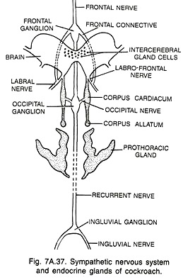
iii. Sympathetic Nervous System:
It comprises the following ganglia and nerves:
(i) Frontal ganglion:
It lies on the dorsal wall of the pharynx in front of the brain. It is connected with the hind brain by the frontal connectives. A frontal nerve arises from the frontal ganglion anteriorly to the frontal region. The recurrent (visceral) nerve runs from the frontal ganglion posteriorly above the oesophagus.
(ii) Occipital ganglion:
After running a short distance, the recurrent nerve becomes thickened to form the occipital ganglion. Three nerves arise from the occipital ganglion. The two lateral ones are called occipital nerves: each runs to the corpus cardiacum (Endocrine gland) of its side. The median one, the recurrent nerve passes posteriorly on the oesophagus and forms ingluvial ganglion near the crop.
(iii) Ingluvial ganglion:
As mentioned above, the median nerve of the occipital ganglion becomes thick posteriorly and gives rise to the ingluvial ganglion near the crop. A pair of lateral ingluvial nerves arises from the ingluvial ganglion posteriorly over the crop.
Essay # 12. Endocrine System of Cockroach:
It consists of:
i. Inter-Cerebral Gland Cells:
They are situated on the brain and secrete brain hormone which activates the prothoracic glands to secrete their hormone.
ii. Corpora Cardiaca:
These rods shaped paired structures lie on the sides of the oesophagus behind the brain. Corpora cardiaca secrete a growth hormone.
iii. Corpora Allata:
These small rounded paired structures are situated just behind the corpora cardiaca. In the nymph corpora allata secrete a juvenile hormone (= neotinin), which retains the nymphal characters and checks the appearance of adult characters.
When the juvenile hormone is absent, it permits the appearance of the adult characters. It is important to note that corpora allata again becomes active in adult cockroach and secrete a gonadotropic hormone, which regulates the development and functioning of the reproductive organs.
iv. Prothoracic Glands:
These large irregular paired glands are located in the pro-thorax and secrete a hormone, the ecdyson to control ecdysis (moulting of the body wall) of the nymph. These glands degenerate after metamorphosis.
Essay # 13. Sense Organs of Cockroach:
Cockroach has several types of sense organs (= receptors). Except the photoreceptors (= eyes), all other receptors, occur in the skin and are called sensillae. The sensillae occur in the epidermis of the body wall. A sensilla has a modified sensory cell innervated by a nerve fibre, trichogen cell and tormogen cell. The secretion of trichogen cells form bristles and spines.
The tormogen cell forms socket. Sensillae are of the following types:
(i) Tactile Sensillae:
They occur all over the body, but are more abundant on the antennae, tibiae of legs and cerci. They respond to touch.
(ii) Gustatory Sensillae:
The sensillae for taste occur on the tips of the maxillary and labial palps and on the epipharynx.
(iii) Olfactory Sensillae:
The sensillae for smell are mainly found on the antennae, and maxillary and labial palps.
(iv) Auditory Sensillae:
The sensillae for hearing are located on the anal cerci.
The antennae, perhaps also bear sensillae for humidity and temperature.
Essay # 14. Compound Eyes of Cockroach:
The two compound eyes occupy a large area on each side of the head. Each eye is black, kidney shaped and bears about 2000 facets externally. The facets are transparent and more or less hexagonal biconvex areas. Each facet actually represents a visual element, the ommatidium. Thus, each compound eye contains about 2000 ommatidia.
An ommatidium consists of two parts: diopteric and receptive. The diopteric part is cone is secreted by four cone cells. Cone cells are surrounded by iris pigment sheath. The receptive part is made up of spindle shaped rhabdome secreted by eight retinular cells or retinulae.
The inner end of each retinular cell is continued into a nerve fibre, the axon which pierces through the basement membrane and joins the optic nerve. The retinular pigment sheath surrounds the region of retinular cells.
The rays which enter obliquely fall on the pigment sheaths and are absorbed while rays of light which enter ommatidia parallel to their long axis help in the formation of image. Thus each ommatidium sees only a small part of object, therefore, the image formed by the eye as a whole consists of several pieces which are put together to make up the whole picture received by the eye.
Such an image is called mosaic image and the eye is said to have mosaic vision. The ocellae or fenestrae or ocellar spots located near the compound eyes are regarded undeveloped simple eyes by some and functional by others because they are sensitive to light.
Essay # 15. Reproductive System of Cockroach:
The cockroaches are dioecious (unisexual) animals. They exhibit sexual dimorphism, e., male and female individuals can be distinguished externally. The female cockroach bears broad abdomen, brood pouch, but lacks anal styles, as present in the males.
Male Reproductive System (Fig. 7A.41):
The reproductive system of male cockroach consists of the following parts:
i. Testes:
There are two testes which are present in the lateral sides in the 4th to 6th abdominal segments. The testes are prominent in young insects, but get reduced in adults.The sperms produced by the follicles of the testes are transferred to the vas deferens through the vasa efferentia.
ii. Vasa-deferentia:
These are two fine ducts. Each vas deferens arises from each testis and opens into ejaculatory duct through seminal vesicle.
iii. Utricular gland (= Mushroom shaped gland):
The junction of the vasa deferentia and the ejaculatory duct is surrounded by a mushroom-shaped gland, utricular eland lying in the 6th-7th abdominal segments. It consists of long tubules, small tubules and seminal vesicles. Long tubules are present at the periphery. Their secretion forms the innermost layer of spermatophore.
Small tubules provide nourishment to the sperms Spermatophore is a pear-shaped capsule, about 1.5 mm long, having a three-layered wall and containing spermatic fluid. The seminal vesicles are present on the ventral surface of the anterior part of the ejaculatory duct. They are numerous small sacs which store the sperms.
iv. Ejaculatory duct:
The base of the utricular gland leads into an elongated muscular tube, the ejaculatory duct. The latter opens to the outside by an opening, the male genital pore lying close to ventral phallomere. The secretion of ejaculatory duct forms the middle layer of spermatophore. The spermatophores are passed outside through the gonopore.
v. Phallic gland (= Conglobate gland):
It is a large elongated sac-like structure which lies beneath the utricular gland and the ejaculatory duct. It tapers posteriorly and terminates into a duct near the male gonopore on the left phallomere. The secretion of phallic gland forms the outer most layer of the spermatophore.
vi. Phallomeres or Male gonapophyses or External genitalia:
There are three chitinous structures that surround the male gonophore. These are right, left and ventral Рhallomeres.
(i) The right phallomere:
Its front part is formed of two opposing plates and the hind part consists of curved serrate lobe which bears two large and pointed teeth like processes at its free end, several small teeth like processes along its border and a hook at its basal part.
(ii) The left phallomere:
It consists of four parts:
(a) Titillator:
It is on left side and bears a hook,
(b) Pseudopenis:
It has a swollen bulb-like apex,
(c) Asperate lobe:
It bears the opening of phallic gland,
(d) Accutolobus:
It is on the left side and bears a terminal hook.
(iii) The ventral phallomere:
It consists of a broad plate and bears the male gonophore. The phallomeres are helpful in the transfer of the spermatophore from male cockroach to female cockroach and their some structures are used to open the female genital chamber.
The sperms produced by the sperm follicles of the testes are passed to the vasa- efferentia which refer to the vasa-deferentia. The vasa-deferentia transfer them to seminal vesicles where they are stored for some time. The secretion of peripheral tubules forms the inner most layer of the spermatophore. Small tubules provide nourishment to the sperms.
Middle layer of the spermatophore is formed by the secretion of the ejaculatory duct. The secretion of phallic gland forms the outer most layer of the spermatophore. Three layered spermatophores are transferred to the female genital chamber by the male phallomeres.
Female Reproductive System:
It consists of the following organs:
i. Ovaries:
There are two yellowish ovaries, one on either side of the alimentary canal. They are embedded in the fat bodies from the 2nd to the 6th abdominal segments. Each ovary consists of eight ovarioles or ovarian tubules which produce ova.
All the filaments of the eight ovarioles of each ovary are united to form a ligament and the ligaments of both ovaries meet in the middle line and get attached to the fat bodies. Eggs of cockroach are centrolecithal (yolk is located at the centre).
ii. Oviducts:
All the eight ovarioles of each ovary unite to form a short tubular and muscular structure, known as oviduct. Each oviduct receives ova from the ovarioles of its side and passes them to the next organ, the common oviduct.
iii. Common Oviduct (= Vagina):
The two oviducts run posteriorly and unite to form a short wide common oviduct. The latter opens into the genital chamber by a vertical slit, the female genital pore.
iv. Genital pouch:
It encloses a boat shaped cavity. The front part of this cavity lies close to the female genital pore and hence called the genital chamber, while the posterior part is called vestibulum or oothecal chamber, because during the breeding it contains ootheca, a structure which contains 14-16 eggs (fertilized ova).
v. Spermathecae:
These are a pair of sacs of unequal size, lying in the 6th abdominal segment. Left one is larger and stores the sperms received from the male during copulation. Right sperm theca is non-functional. Both the spermathecae open by a common duct into the genital chamber.
vi. Colleterial glands:
They are two very much branched tubuler glands, which are unequal in size and embedded in the fat. The left gland is large. Both the glands open independently on the dorsal side of the genital chamber. The secretion produced by these glands forms the oothecal case of the ootheca.
vii. Gonapophyses:
The six chitinous plates, the phallomeres, surrounding the female genital pore, are termed as gonapophyses. There is a pair of anterior gonapophyses and two pairs of posterior gonapophyses which function as ovipositors. The latter are used to carry eggs to the oothecal chamber.
The male and female cockroaches come together by their posterior ends and with the help of the male phallomeres, the spermatophores are transferred to the genital chamber of the female where the sperms are liberated from the spermatophores and reach the left spermatheca slowly.
The eggs come from both the ovaries alternately into the common oviduct and pass through the female gential pore into the genital chamber where they are fertilized by the sperms coming from the left spermatheca. The secretion of collaterial glands forms the egg case of the ootheca. The ootheca is placed by female cockroach in dark and warm region.
The placement of ootheca is called oviposition. On an average, a female produces 9-10 oothecae, each containing 14-16 fertilized eggs in two rows. Thus 14-16 young cockroaches (nymphs) develop in one ootheca. The nymph grows by moulting about 13 times to reach the adult form.
There is paurometabolous (gradual) metamorphosis in cockroach. A stage in the development of an insect between two moults is called instar. The interval between two successive moults of an insect or other arthropod is known as stadium.
Essay # 16. Interaction of Cockroach with Mankind:
Cockroaches cause damage to the household materials such as clothes, purses, shoes, etc. They also eat and destroy human food such as bread, fruits, cheese, etc. Since they also live in sewage pipes and gutter holes, they carry harmful germs of diseases like diarrhoea, cholera, typhoid, tuberculosis, etc.
They also produce obnoxious smell in kitchens and stores. In South American countries and in Myanmar people eat cockroaches. Many animals such as amphibians {e.g., frogs, toads), lizards, birds and rodents eat cockroaches. Thus they are the part of food chain. Cockroaches are used as experimental animals as they can be obtained easily without causing much ecological imbalance.


