
The following points highlight the top eight types of classical endocrine gland. The types are: 1. Hypothalamus 2. Pituitary Gland 3. Pineal Gland 4. Endocrine Pancreas 5. Thyroid Gland 6. Parathyroid Gland 7. Thymus Gland 8. Adrenal Gland.
Contents
- Classical Endocrine Glands: Type # 1. Hypothalamus:
- Classical Endocrine Glands: Type # 2. Pituitary Gland:
- Classical Endocrine Gland: Type # 3. Pineal Gland:
- Classical Endocrine Glands: Type # 4. Endocrine Pancreas:
- Classical Endocrine Gland: Type # 5. Thyroid Gland:
- Classical Endocrine Glands: Type # 6. Parathyroid Gland:
- Classical Endocrine Glands: Type # 7. Thymus Gland:
- Classical Endocrine Glands: Type # 8. Adrenal Gland:
Classical Endocrine Glands: Type # 1. Hypothalamus:
The hypothalamus is anatomically basal part of diencephalon. It forms the walls and lower part of the third ventricle of brain. In mammals, neurosecretory cells are highly aggregated in this region. It controls the entire endocrine mechanism within the body through pituitary gland.
Hence it can be popularly called as super master gland. It secretes various releasing hormones which in turn stimulate specific cells of pituitary for secretion of pituitary hormones.
Anatomy of Hypothalamus:
Hypothalamus lies in close contact with optic chiasma, the infundibulum, median eminence and mammillary body. The vascular link between the median eminence and the pituitary gland is known as the hypothalamohypophysial portal system (Fig. 4.1).

Histology of Hypothalamus:
Within the hypothalamus, huge clusters of neurosecretory cells present called hypothalamic nuclei which are located around the third ventricle.
In mammals, hypothalamus consists of paired neurosecretory hypothalamic nuclei like anterior hypothalamic (AH), suprachiasmatic (SC), ventromedial (VM) dorsomedial (DM), posterior hypothalamic (PH), supra optic (SO), Para-ventricular (PV), Periventricular and actuate nuclei (AN).
These nuclei are responsible for secretion of different releasing hormones from hypothalamus. The hypothalamic hormone secretion is controlled by neural activity or feedback mechanism (Fig. 4.2, 4.3 & 4.4).


Hypothalamic hormones (see Table 4.1) go from the neurons to the blood capillaries of the portal system and then to the target cells of the pituitary.

Classical Endocrine Glands: Type # 2. Pituitary Gland:
In vertebrates, pituitary gland is known as ‘Master gland’, because it initiates all the physiological functions, as well as controls the secretions of other endocrine glands. This gland was identified by Vesalius in the 16th century. After that its biological functions worked out by Aschner (1910), Smith (1926), Riddle (1933) etc. Holmes and Ball (1974) studied the ultra-structural details.
The name pituitary is derived from Latin word “Pituita” means mucus.
Development of Pituitary Gland:
This gland is developed from two embryological primordia. The posterior pituitary (neurohypophysis) is derived from infundibular region of diencephalon and anterior pituitary (adenohypophysis) is developed from the buccal epithelium.
Buccal epithelium gives an evagination, called Rathke’s pouch. At the sometime, infundibulum shows downward projection towards Rathke’s pouch from the floor of diencephalon. Ultimately both the walls fuse together. Thereafter Rathke’s pouch losses its connection from the surface epithelium. Cells of the Rathke’s pouch proliferate to give rise to the histological details of anterior pituitary.
Accordingly nerve fibres grow into the infundibulum from hypothalamus and give rise the neurosecretory cells of posterior pituitary. Continued proliferation of cells of Rathke’s pouch leads to the reduction of lumen of the pouch to a residual cleft or inter-glandular cleft.
Infundibular growth finally forms the stalk of pituitary gland, by means of which the gland is connected with the diencephalon. Thus the gland remains under the hypothalamus, hence it is known as hypophysis. It is connected with the 3rd ventricle of diencephalon. This gland is placed in a concavity of sphenoid bone called sellaturcica (Fig. 4.5).

External Anatomy of Pituitary Gland:
1. The pituitary gland is very small and reddish or pinkish in colour.
2. Pituitary Gland is more or less oval in shape.
3. Sizes vary from 2-10 µm in length and 0.5-0.7 mm in thickness.
4. Anatomically it is divided into two parts—Adenohypophysis and neurohypophysis.
5. Pituitary gland is innervated by sympathetic nerves and blood vessels (Fig. 4.6).

Description of Adenohypophysis:
Adenohypophysis in most important part of pituitary and it constitutes about 75% of the gland. It is glandular in nature and secretes various kinds of hormones. Histologically it is divided into three parts—pars distal is, pars inter-media and pars tuberalis.
(A) Pars Distalis:
1. It is the main part of adenohypophysis.
2. This part of the gland is composed of irregular masses and cords of epithelial cells separated by sinusoids and supported by loose framework of connective tissue.
3. Routine staining of pars distalis with orange-G, PAS, Mallory Aniline blue etc. reveals two major varieties of gland cells like chromophobe cells and chromophil cells which constitute about 60% of glandular part of Pars distalis (Fig. 4.7).

(a) Chromophobe cells:
These types of cells are not stained with any dye. These cells have less cytoplasmic and lack secretory granules. E.M. structure shows that chromophobe cells contain very fine basophilic granules. Some histologists believe that these cells are embryonic condition of chromophil cells.
(b) Chromophil cells:
These types of cells take up both acid and basic dye. Thus these cells are further divided into acidophil and basophil cells.
Among these acidophil cells are about 85% and basophil cells are about 15%.
These cells secrete different type’s hormones.
Following types of chromophil cells are identified:
1. Acidophil cells: It is of two types (a) STH cells (b) LTH cell
(a) STH cells:
These cells ovoid or round in shape. Nucleus is central in position and cytoplasm is packed with acidophil granules. Size of the granules varies from 350-400 mµ. Histo-chemically orange-G positive. It secretes STH hormone which is protein in nature.
(b) LTH cell:
These cells are ovoid or ellipsoidal in shape. Nucleus is at the centre and cytoplasm is packed with large granules. Sizes vary from 600- 900 mµ. Histo-chemically orange-G positive. It secretes protein hormone prolactin or LTH.
2. Basophil cells:
These cells are of four types: (a) FSH cells (b) LH/ICSH cells (c) TSH cells (d) ACTH cells.
(a) FSH cells:
These cells are large, round in shape. The nucleus is slightly wrinkled and the cytoplasm is packed with dense spherical granules. Sizes vary from 150-300 mµ. These cells secrete hormone FSH which is glycoprotein in nature. Histologically orange-G negative.
(b) LH/ICSH cells:
These cells are round or polygonal in shape. Nucleus is wrinkled. Size of the cytoplasmic granules vary from 100-300 mµ. These cells secrete glycoprotein hormone, LH in case of female and ICSH in males. Cells are histo-chemically orange-G negative.
(c) TSH cells:
These cells are irregular in shape and nucleus is round and centrally placed. Cytoplasmic granules are fine and measured about 100-140 mµ. These cells secrete glycoprotein hormone TSH. It is orange- G negative.
(d) ACTH cells:
These cells are round and small in sizes with central round nucleus cytoplasmic granules are very fine and comparatively low in number and measured about 100 mµ. They secrete glycoprotein hormone ACTH. Histo-chemically they are orange-G negative (Fig. 4.8) & (Table 4.2).


(B) Pars Inter-media:
This zone is anatomically associated with the neural lope and separated from pars distal is by inter-glandular cleft.
Anatomical variations:
1. It is absent in whales and dolphin;
2. In desert mammals it is well developed.
3. In man pars inter-media is reduced.
Histology of Pituitary Gland:
It consists of basophilic and chromo-phobic cells. It consists of melanocyte cells and fibrous tissue. E.M. study reveals that nucleus of melanocyte is round and central in position. Several mitochondria, ER, Golgi bodies are well apparent. Cytoplasm is filled up with secretory granules (Fig. 4.9).

(C) Pars Tuberalis:
It lies adjacent to pars distalis and pars inter-media. It is very close to the infundibulum stalk. It is separated from infundibulum by fibrous connective tissue. The main component of pars tuberalis in cuboidal or short columnar cells. These cells contain lipid and glycogen droplets. Hence they are Sudan black-B and PAS “+”ve. Actual endocrine function is not clear.
Description of Neurohypophysis:
Neurohypophysis is nervous in origin. Anatomically it constitutes three parts—pars nervosa, median eminence and infundibulum.
Histology:
(A) Pars nervosa:
It is the main component and histologically following elements are found:
1. Pituicytes:
It is a type of neurosecretory cell. Cell is slightly elongated with distinct cell body. Nucleus is round oval in shape with prominent chromatin materials. Dendritic processes are absent. Axonic process is slightly elongated and terminal portion is comb like in appearance.
Mitochondria, ER, Golgi apparatus etc. are well apparent. Secretory vesicles and yellowish brown pigments are scatteredly distributed within the axonic process. Cells are Bromophenol Blue positive.

2. Mast cells:
These are round cells with central nucleus.
3. Herring bodies:
Non-nucleated nerve fibres aggregate together to form herring bodies.
4. Neuroglial cells:
Polygonal or round neuroglial cells or distinct between the pituicytes.
5. Nerve and sinusoids:
These are well apparent.
(B) Infundibulum:
It is the stalk of pituitary. It is made up of neuroglial cells. Blood sinusoids and few neurosecretory cells present.
(C) Median eminence:
It is made up of hypothalamic cells neuroglia and blood vessels.

Classical Endocrine Gland: Type # 3. Pineal Gland:
Pineal gland is an important endocrine gland in case of higher vertebrates. The name is derived from its pinecone like structure. This gland receives its stimuli through vision, hence it is also known as third eye. This gland was first discovered by Herophilus about 2000 yrs. ago.
Decartes (1646) said that it was the “seat of soul” and receives stimuli through eye. Lamer (1948) first established the biological function of this gland. He administrated pineal extract of cow into the amphibian skin and showed bleaching action on the skin.
Development of Pineal Gland:
Pineal gland is embryo- logically derived from neuroectoderm of the di-encephalic roof. The gland remains its connection with the diencephalon by a thin stalk.
Anatomy of Pineal Gland:
1. This gland is located on the dorsal surface of the brain (Fig. 4.11).

2. It is embedded in between the cerebellum and cerebrum.
3. The shape is flattened stalk like structure hence it is called epiphysis (Fig. 4.12).

4. It has sympathetic innervation.
Histology of Pineal Gland:
Pineal gland consists of two types of major cells—Pinealocytes and Astrocytes. Besides these connective tissue cells, mast cells, nerve fibres etc. also present.
EM Structure of Pinealocytes (Fig. 4.13)

The following points highlight the main features of Pinealocytes:
Features:
1. It is a neuro secretory cell.
2. It has nucleated cell body like structure and also possess short and long processes like neuron.
3. It is surrounded by plasma-membrane which is lipoprotein in nature.
4. Nucleus is slightly irregular in shape. It has prominent nucleolus and little RNA.
5. GER shows peripheral localization whereas SER are apparent near nucleus.
6. Free ribosomes are distributed in the cytoplasm.
7. Golgi apparatus are prominent with vesicles and measure about 40-100 nm.
8. Mitochondria are very long (4-5.8 µm) and has elongated tubular cristae.
9. Centrioles are absent.
10. Lipid droplets and electron dense vesicles are apparent in the cytoplasm.
11. Dendrite like two short processes present.
12. Axon like long process has comb like ending.
13. Parallely arranged electron dense microtubules present and is known as synaptic ribbon. Each tubule has electron dense core about 120 nm diameter.
Types:
There are two types of pinealocytes—
1. Light pinealocytes:
These are many and large cell types. Nucleus is greatly lobulated. Cytoplasm is basophilic and is devoid of glycogen. SER are more in numbers in these cells. Ribosomes are more prominent. These cells are chiefly found near capillary endothelium. Synaptic ribbon is prominent.
2. Dark pinealocytes:
These are few in number and comparatively small in sizes. Nucleus is comparatively less lobulated. Large amount of glycogen droplets present in the cytoplasm. These are distributed near synaptic neurons.
EM Structure of Astrocytes (Fig. 4.14)

1. Present in between the pinealocytes and
2. These are small stellate shaped cells and are supposed to be modified neuroglial cells.
3. Nucleus is elongated and dense in nature.
4. Corpora arenacea
5. Have few mitochondria, Golgi bodies, ER and ribosomes.
6. Occasional deposition of glycogen droplets.
Note:
In human, pineal body often contains corpora arenacea, which are extracellular mineralized concentric organisation and mainly consist of calcium phosphate and carbonates. Function is not clear (wood, 1981) (Fig. 4.15).

Classical Endocrine Glands: Type # 4. Endocrine Pancreas:
Pancreas is a very important gland in vertebrates. This gland contains both exocrine and endocrine parts hence it is called as Mixocrine gland. The endocrine part was first described by Langerhans in 1869, hence it is popularly known as Islets of Langerhans. In mammals, this gland is well developed.
Development of Endocrine Pancreas:
Pancreas is derived from endodermal lining of the duodenum.
Anatomy of Endocrine Pancreas:
1. The gland is elongated leafy structure lies in between the duodenal loop and is connected with the intestinal wall by pancreatic duct (Fig. 4.16).

2. The gland is creamy white or light pink in colour.
3. The gland can be divided into a head, body and tail.
4. The head is an expanded portion that lies in the C-shaped curve of duodenum. It is joined to the duodenum by connective tissue.
5. The centrally located body and tapering tail extend to the left.
6. The pancreatic duct extends through the length of the gland and empties into the duodenum.
7. It is well vascularized and innervated by nerve fibres.
Histology of Endocrine Pancreas:
1. The gland is invested by thin fibrous tissue.
2. Internally consists of exocrine and endocrine components and the ratio is 85:15.
3. Exocrine units or acini are basophilic in nature. Each acinus is lined by simple epithelium and the lumen of acinus is very narrow. The acinal cell contains basophilic zymogen granules in the cytoplasm.
4. Islets of Langerhans are the endocrine units and are scatteredly distributed among the acini.
5. Islets are well vascularized mass of cells.
6. Shapes of islets vary and sizes vary from 20-300 µn.
7. Islets are whitish in colour and internally consist of blood sinusoids. Number of islets varies in species to species, for instance in man, there are about 1 million islets (Fig. 4.1 7).

8. After light microscopy, three types of cells are identified after Mallory Azan method—

9. Electron microscopy reveals that islets cells are of two categories—
(a) Major cells:
Three types, α, β and δ cells.
(i) α-cell:
Cells are tall and cylindrical. Cytoplasmic granules are about 250 nm in diameter. Each granule is invested by lipoprotein membrane. Granule is oval is shape and bears light eccentric cone. Nucleus is lobulated type. Cytoplasm contains Golgi complex, ER, ribosomes and filamentous mitochondria (Fig. 4.19).

(ii) β-cell:
Cells are more or less round or irregular in shape with central round nucleus. The cytoplasmic granules are hexagonal and lined by double layered membrane. Granules are about 300 nm in diameter with dense crystalline core. Cells contain more ER and free ribosomes than a-cells. Golgi bodies and mitochondria are apparent (Fig. 4.20).

(iii) δ-cell:
Cells are large spindle shaped. Cytoplasmic granules are very large and are about 325 nm in diameter. Each granule is lined by smooth membrane and contains homogeneous matrix. Few glycogen and lipid droplets are visible. Cytoplasmic inclusions like ER, ribosomes, mitochondria, Golgi bodies are apparent. Nucleus is elliptical in shape (Fig. 4.21).

(b) Minor cells: Three types
(i) PP cells:
These cells are found in between the duct of acini. They secrete pancreatic polypeptide.
a. It inhibits bile secretion
b. It inhibits enzyme secretion
c. Inhibits HCO–3 secretion.
(ii) Di cells:
Cells are columnar type and granules are 1.18 nm in diameter. Scatteredly distributed and secrete vasoactive intestinal polypeptide.
a. It increases enzyme secretion.
b. Increases bile secretion.
(iii) Ec cells:
Secrete exo-chromaffin hormone.
Remark:
In mammals, pancreatic islets are called β-islets, because of more in numbers of β-cells (Fig. 4.18).

Classical Endocrine Gland: Type # 5. Thyroid Gland:
The thyroid gland is one of the important endocrine gland in vertebrate series. Its hormone is important in tissue differentiation, metamorphosis and iodine metabolism. From evolutionary standpoint, through iodine binding capacity it is believed that thyroid gland is evolved from the endostyle of protochordates.
Kendall (1918) isolated the active component of the thyroid called thyroxine. The thyroid is unique among vertebrate endocrine glands in that it stores its hormones extracellularly.
Development of Thyroid Gland:
Thyroid gland develops from the endoderm of the cephalic portion of alimentary canal of embryo. A sac like diverticulum first appears in the mid-ventral surface of the pharynx at the level of 2nd pair of pharyngeal pouches. The diverticulum expands and remains attached with the pharynx by a narrow solid duct, called thy- roglossal stalk. Later the stalk atrophies and the gland remains free (Fig. 4.22).

Anatomy of Thyroid Gland:
In mammals, thyroid gland is lobed in nature and lies on either side of up per part of trachea, just below the cricothyroid cartilage. In man both the lobes are connected by Isthmus Bridge. The gland is oval or pear shaped. The gland is light pink in colour and is covered by fibrous capsule.
The size of the gland varies in species to species. For instance, in human, the gland measures about 2.5-4 cm in length and 1.5-2 cm in width and 1-1.5 cm in thickness. Normal weight of the gland in human is 15-40 gm. It is well vascularized by thyroid artery and is innervated by sympathetic nerve fibres from cervical region of spinal cord (Fig. 4.23).

Histology of Thyroid Gland:
On light microscopy:
The gland is invested by outer capsule which is made up of white fibrous connective tissue. Internally it consists of many thyroid follicles. For instance, in rat there are about 100,000 thyroid follicles. The follicles are of various sizes which vary from 15-150 µm in diameter. Large follicles are usually distributed towards the periphery and smaller are present near the centre.
Each follicle is lined by simple epithelium and epithelium is made up of cuboidal cells. Very thin basement membrane present outside the follicular epithelium. The lumen of the follicle is filled up with homogeneous, gelatinous material which is eosinophilic in nature after H-E stain.
Colloid is protein in nature is secreted by epithelial cells. Thyroid follicles are separated by connective tissue. Within the meshes of connective tissue few lymphocytes, histocytes, sections of blood vessels and nerves present (Fig. 4.24).

On Electron microscopy:
Electron microscope reveals that follicular cells contain Golgi apparatus near the luminal end. Mitochondria, ER and lysosomes are scattered throughout the cytoplasm. Nucleus is well apparent at the centre of the cell. There are fine terminal bars at the apical side of the follicular cells. Cells are held together with the help of terminal bars.
Histo-chemically lipid droplets, different proteins laden granules are observed. There are two types of cell are found chief follicular cells and Para follicular cells or C-cells. Chief follicular cells are the main constituent of follicular epithelium. Para follicular cells may occur within the follicular epithelium or in the regions between or adjacent to the follicles (Fig. 4.25).

In this article we will discuss about Comparative account of chief follicular cells and Para follicular Chief follicular cells. After reading this article you will learn about: 1.Chief follicular cells 2.Para follicular cell
Chief follicular cell:
1. Luminal side bears microvilli
2. Comparatively large in number.
3. Comparatively less number of mitochondria.
4. Has less oxidative enzyme.
5. Nucleus is round.
6. Enlargement of Golgi apparatus from TSH treatment
7. Cytology not altered by high blood calcium level.
8. ER larger in diameter.
9. Secretes T3 and T4.
10. Binds antibody to thyroglobulin.
Para follicular cell:
1. Microvilli absent.
2. Comparatively small in number.
3. Comparatively mitochondrial rich.
4. Has high concentration of oxidative enzyme.
5. Nucleus is irregular in outline.
6. Golgi apparatus prominent.
7. Degranulation due to high blood calcium level.
8. Comparatively less in diameter.
9. Secretes calcitonin.
10. Binds antibody to calcitonin
Classical Endocrine Glands: Type # 6. Parathyroid Gland:
Parathyroid gland is anatomically associated with thyroid gland. It is responsible for calcium and phosphorus metabolism. The first anatomic description of this gland was given by Sand storm (1880).
Development of Parathyroid Gland :
Parathyroid gland is originated from pharyngeal endoderm in association with 3rd and 4th pharyngeal pouches of mammals.
Anatomy of Parathyroid Gland:
1. In mammals, parathyroid glands are well developed and are embedded in the posterior surface of the thyroid gland.
2. Usually there are two parathyroids but four parathyroids are found in man, dog, cat, rabbit etc.
3. In man, four parathyroid are small oval bodies (6 x 3 x 2 mm) embedded in the thyroid tissue. Total weight is about 140 mg.
4. The gland is well vascularized by superior and inferior thyroid artery.
5. The gland is innervated by sympathetic nervous system (Fig. 4.26).

Histology of Parathyroid Gland:
1. Histologically the gland is made up of epithelial cells.
2. Two types of cells — Chief cells and oxyphil cells.
3. Chief cells are more numerous and comprise about 99% of the Para thyroid’s cellular population cells are cuboidal in nature. The nucleus is round and centrally placed. Cytoplasm contains Golgi complex, ER, mitochondria and cytoplasmic granules. Chief cells posses β-androgenic receptors as well as glucocorticoid receptors.
4. Oxyphil cells are large polyhedral in nature. These cells represent only about 1 % of the total parathyroid cellular population. These cells are eosinophilic cells rich in mitochondria.
5. Small colloidal vesicles and variable amounts of adipose tissue may be observed within the gland (Fig. 4.27).

Classical Endocrine Glands: Type # 7. Thymus Gland:
Thymus is an important endocrine gland in mammals and plays an important role in immune mechanism at a young stage. It is relatively large organ at the time of birth and the maximum weight is attained in young age. For instance, in human children, the weight is about 30-40 gms. It undergoes age involution and is partially replaced by fat and connective tissue.
Miller (1961) first recognized the immunological function of thymus. Good (1964), Cooper (1976), Londry et al. (1989), Kendeu (1990), Ritter and Crispe (1992) etc. studied the histology and endocrine functions of this gland.
Development of Thymus Gland:
Thymus gland arises as a thickening of the endodermal lining of pharyngeal pouches on each side. The thymic masses subsequently become separated from the pouches and remain as a separate gland. In mammals 3rd and 4th pouches are the sole contributors.
Morphological Description of Thymus Gland:
This gland lies between the posterior part of the thyroid gland and anterior end of pericardium. The gland lies on the surface of heart. It is bi- lobed structure. Each lobe is elongated flask shaped. The gland is well vascularized by blood vessels and lymph vessels. It is innervated by vagus nerve and cervical sympathetic nerves (Fig. 4.28).

Histology of Thymus Gland:
This gland is invested by a thin collagen connective tissue capsule. From the capsule connective tissue septa enter the gland and helps in the development of thymic lobules. Each lobule is of 0.5-2 mm in diameter. Each lobule comprises of dark stained peripheral zone, called cortex and central pale zone, called medulla. Within the gland, septa and trabeculae are formed by the connective tissue capsule extensions (Fig. 4.29).

(A) Cortex:
1. It is made up of loosely packed mass of cells. Cells are of different types.
2. The reticular cells are stellate type and have protoplasmic processes. The cells are acidophilic in nature, having oval nucleus (7-11 µm in diameters) with one or two nucleoli. EM study reveals that the processes are joined by desmosomes. The cytoplasm certain mitochondria, ER ribosomes, Golgi apparatus. Few vesicles are found, that are supposed to be lysosomes (Fig. 4.31).

3. Thymocytes, they are of large and small type. These cells are supposed to be modified lymphocytes. Cortex is constituted by about 80% thymocytes.
Small thymocytes are more abundant than large thymocytes. Small thymocytes have dense round nucleus. Cytoplasm with few ribosomes, mitochondria and GER. Small type is about 7µm in diameter.
Large thymocytes have oval nucleus with rich chromatin material. Cytoplasm contains polyribosomes, few mitochondria, GER, vesicles and lipid droplets. They are more than 11µm in diameter.
4. A few plasma cells present.
5. Macrophages are scatteredly distributed in the cortex.
6. At the periphery of cortex and near the blood capillaries, the protoplasmic processes of reticulocytes form a continuous thin partition to separate the parenchyma from blood capillaries, called blood-thymus barrier.
(B) Medulla:
1. It is the light stained central area of lobule.
2. Medulla is filled up with reticular cells, few thymocytes mast cells, eosinophil’s and very few macrophages.
3. Reticulocytes are of various shapes. Some have small cytoplasmic or large cytoplasmic filaments.
4. Small type thymocytes present.
5. Macrophages are rarely present.
6. Plasma cells are absent.
7. In the centre of medulla thymic or Hassal’s corpuscles present, which are concentrically arranged layers of modified flattened reticular cells (Fig. 4.30).

Each corpuscle has a central calcified cell. Reticular cells are attached by desmosomes. Corpuscles are 100 µm or more in diameter. Each corpuscle in a section looks somewhat like a slice of onion.
Classical Endocrine Glands: Type # 8. Adrenal Gland:
Adrenal gland is well known important endocrine gland in vertebrate series. Anatomically it is well developed in mammals. It performs various physiological functions. Eustachius (1563) first described adrenal gland in man. Addison (1855) first determined the clinical significance of the gland.
Development of Adrenal Gland:
Cuvier (1805) recognised two distinct regions in mammalian adrenal— cortex and medulla. Two components of this gland originate from different embryonic primordia. The cortex is derived from lateral mesoderm in close association with gonads.
The medulla differentiates from neural crest cells along with sympathetic ganglia. Hence cortical secretion is controlled by pituitary whereas medullary secretion is controlled by nervous stimulus.
Location of Adrenal Gland:
This gland is situated above the kidney hence it is known as supra renal gland (Fig. 4.32).

Anatomy of Adrenal Gland:
Light microscopic study:
The gland is oval or slightly triangular in nature. It is outerly covered by fibrous capsule. Histologically it consists of outer cortex and inner medulla (Fig. 4.33).

Cortex:
1. Cortex is made up of epithelial cells and is provided with blood sinusoids and nerves in section.
2. Cortex is histologically subdivided into 3 zones—zona glomerulosa, zona fasciculata and zona reticularis.
3. Zona glomerulosa:
This is the thin outer zone lies in parallel with outer fibrous capsule. It is made up of group of small columnar cells. These cells are arranged in clusters like bunch of grapes. This zone is light bluish in colour after H-E stain due to presence of basophilic granules.
The zona glomerulosa is not apparent in all mammals, like mice, lemurs and monkeys.
4. Zona fasciculata:
It is the widest middle layer of cortex. It is formed by polyhedral cells. Cells are arranged in the form of narrow columns or cords. Nuclei are larger and less dense. The cords of cells are surrounded blood sinusoids. Cytoplasm contains brownish yellow pigment. After H-E stain, cytoplasm shows light pink in colour.
5. Zona reticularis:
It is the terminal sub-layer of cortex. It is made up of flattened irregular cells. Cells are arranged in irregular network fashion, this zone also contains few reticulin fibres. Hence this zone is called reticularis. Nuclei are dense in colour. After H-E stain, this zone looks much dark pink than fasciculata zone.
Medulla:
1. Medulla consists of large irregular cells.
2. Cells are packed with cytoplasmic granules.
3. Large distinct nucleus present in each cell.
4. Large blood sinusoids are well apparent.
5. After H-E stain, medullary cells appear bluish in colour.
6. Medullary cells are also known as chromaffin cells, because treatment of these cells with potassium dichromate or chromic acid results in formation of yellowish or brown oxidation product, the chromaffin reaction.
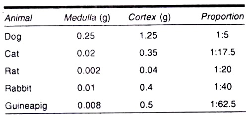
The relative amounts of adrenocortical and medullary tissues vary in different species:
Additional zones:
Several unique adrenocortical zones are known in certain species of mammals. These zones may be conspicuous only at certain times in the life of an animal or in only one sex.
1. The Fetal zone:
In primates, a very con spicuous zone occupies the bulk of the adrenal gland prior to birth. Following birth this zone degenerates rapidly.
2. The mouse X-zone:
The cortex of the mouse adrenal contains a unique X-zone between zona reticularis and the medulla. The X-zone degenerates in males at puberty and in females of most nurse during the first pregnancy.
3. The special zone:
One marsupial brush tailed opossum (Trichosurus vulpecula) possesses a large inner special zone only in adult female.
Electron microscopic study (Fig. 4.34) Cortex

1. Zona glomerulosa:
(a) Cells are comparatively smaller than other zones.
(b) Plasma membrane shows small microvilli.
(c) Well developed smooth ER present.
(d) Golgi apparatus present in very close association with nucleus.
(e) Rich in mitochondria which possess shelf like cristae.
(f) Few lipid droplets are found in the cytoplasm.
2. Zona fasciculate:
(a) Cells are relatively larger and possesses huge amount of lipid droplets in the cytoplasm.
(b) Golgi complex distinct.
(c) Mitochondria are many and tubular in shape.
(d) ER much more in concentration.
(e) Microvilli are well apparent on the surface membrane.
(f) Few cells are spongy in appearance, (Anderson 1984).
3. Zona reticularis:
(a) Cells are comparatively larger.
(b) Lipid droplets are few and scattered in cytoplasm.
(c) Elongated mitochondria are well apparent.
(d) ER and Golgi apparatus are distinct.
(e) Secretory vesicles present.
(f) Microvilli distinct.
Medulla:
1. Cells have smooth plasma membrane.
2. Microvilli absent.
3. Mitochondria, ER and Golgi apparatus distinct.
4. Cytoplasm contains dark granules each granule is enclosed by smooth lipoprotein membrane. The diameter of the granule is about 200 µm.
5. There are two types of cells are found nor epinephrine cells and epinephrine cells.
6. Granules of nor epinephrine cells are much more electron dense than epinephrine cells.