In this article we will discuss about the gametophytic phase, reproduction and sporophytic phase in the life cycle of pogonatum. This will also help you to draw the life cycle of pogonatum.
Contents
Gametophytic Phase of Pogonatum :
(i) External Features of Gametophyte:
The plants are perennial, erect gametophytes and can be differentiated into two regions:
(a) Rhizome
(b) Rhizoids
(a) Rhizome:
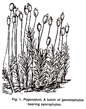
The adult plant has a basal rhizome-like structure which gives rise to many aerial shoots or leafy shoots on the upper side and rhizoids on the lower side. The aerial shoots form thick mat like cushions. (Fig. 1, 2)
(b) Rhizoids:
Rhizome is densely covered with multicellular, thick walled rhizoids with oblique septa. Many rhizoids coil, twist and inter-wine to form rope-like thick structure. Main functions of the rhizoids are anchorage and absorption.
Aerial Shoot: These are erect and arise from the rhizome. It consists of a central axis, the stem which bears scaly and green leaves. Scaly leaves are present on the basal portion of the stem while normal leaves are present on the upper part of the gametophore.
External Structure of the Leaf:
The leaves are costate with long lanceolate lamina and colourless base that en-sheaths the stem.
Each leaf can be differentiated into two parts:
A sheathing base and apical limb.
Sheathing Base:
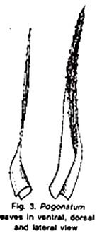
It is colourless, broad membranous structure (Fig. 3). It clasps the stem closely and this provides the external capillary conducting system for water.
Apical Limb:
It is green, narrow and tapering at both the ends (panceolote). It has a thick mid rib region (nerve or costa) and two laterally extended thin and narrow wings or lamina (Fig. 3).
Leaves in Ventral, Dorsal and Lateral View
The transition from sheath to blade is often abrupt with a hinge tissue. This tissue by swelling and shrinking controls outward and inward movement of the blade. Under moist conditions the leaves are divergent exposing the upper surface for maximizing photosynthesis. Under dry conditions the leaves are imbricated reducing water loss.
(ii) Internal Structure of Pogonatum:
1. Rhizome:
A transverse section of rhizome is irregular in outline. It can be differentiated into:
(i) Epidermis:
It is the outermost layer. It is composed of single layer of thick walled cells. In the rhizome, numerous epidermal cells extend to form the rhizoids.
(ii) Cortex:
It is inner to epidermis and is differentiated into two parts:
(a) Outer Cortex.
It is 3-5 layered and is composed of thick walled cells (cells also called stereids).
(b) Inner Cortex:
It is a broad zone of thin walled parenchymatous cells.
The cells of the cortex contain starch. A large number of the leaf traces are also present in the cortex.
(c) Endodermis:
Cortex is separated from central strand by a single layer of enlarged cells interpreted as endodermis.
(d) Central Strand:
It is present in the centre of axis and consists of stereids, hydroids and leptoids cells. Stereids (as a whole being called stereom) are thick walled and constitute the main part of the central strand. Their main function is to provide mechanical support. Hydroids (as a whole being called hydrom) are elongated, thin walled cells.
These are scattered in stereids in groups of 2-3. Their main function is water conduction (similar to xylem of higher plants). Leptoids are present next endodermis and are comparable to the sieve cells of the higher plants.
2. Stem:
Internally the stem of aerial branches shows similar structure with some differences:
(i) Rhizoids absent
(ii) Cortex is made up of only hydroids
(iii) Cortex is encircled by a discontinuous layer of laptoids
(iv) Endodermis absent.
3. Green Leaf:
A transverse section passing through the apical limb can be differentiated into two parts:
(i) Midrib:
It forms the major part of the leaf. It is several layers thick in the centre and gradually merges into the rudimentary wings (lamina) at the margins. On the lower side it has a distinct epidermis which is composed of a single terminal ceils layer of cells.
The central tissue is made of large parenchymatous cells. The hydroids and leptoids are also present in the central tissue of the leaf as scattered cells. On the upper side there is a layer of large cells (Fig. 6A) from which arise many vertical plates of cells called lamellae.
The lamellae constitute the photosynthetic area of the leaf. Each lamella is filamentous, 4-8 cells in height and all the cells, except the terminal one, contain chloroplast.
The terminal cell of the filament is wider, papillose, hyaline and usually larger than the other cells. In P. microstomum the terminal cell splits into two apical cells (Fig. 6B). These lamellae are separated by narrow intercellular spaces which form capillaries to hold water. These spaces also serve for gaseous exchange.
(ii) Wings:
The wings are made only of one layered thick hyaline cells. Lamellae are absent in this region. These wings are absent in this region. These wings may fold over lamellae to prevent water loss.
Reproduction of Pogonatum:
Pogonatum reproduces by two methods:
(i) Vegetative Reproduction:
It takes place by the following methods:
1. By the development of bud-like gemmae on the rhizoids.
2. By primary protonema:
It develops multicellular buds which develops into leafy gametophore.
3. By secondary protonema which may develop from any part of the gametophyte and behave as primary protonema.
4. By fragmentation of rhizome
5. By apsorpy:
Diploid protonema develops from any part of the sporophyte.
(ii) Sexual Reproduction:
It is oogamous. Male reproductive structures are known as antheridia and female reproductive structures are known as archegonia. Majority of the species of Pogonatum are dioecious but P. microstomum is monoecious. These sex organs develop in terminal clusters on the apices of the separate gametophores.
Antheridial Head:
A male plant may have several antheridial heads. The antheridia develop on the tip of the male branches and appear like flower. It is antheridial head (Fig. 7A). It is formed by the perigonial leaves. These leaves are red or orange in colour.
Since antheridia are produced in groups at the base of each perigonial leaf in the position of lateral buds, the apical cell of the stem is not utilised and hence the apical growth of the stem is not affected. The apical growth of shoot continues in the head in following year.
Such growth pattern of the gametophore beyond the antheridial head is known as proliferation. Many sterile, uniseriate, multicellular, hairs like structures are also present in between the antheridia. These are intermingled with antheridia and are called paraphysis (Fig. 7B).
Antheridium:
A mature antheridium can be differentiated into two parts:
Short stalk and club shaped body. The body of the antheridium has a single layer jacket which encloses a mass of biflagellate antherozoids. An operculum is present at the tip of mature antheridium which helps in the liberation of antherozoids (Fig. 7 C, D).
Archegonial Head:
It is the terminal part of the female gametophore. It is formed by the perichaetial leaves and gives a bud like appearance. The archegonia occur in groups of 3-6 and are surrounded by sterile paraphysis and perichaetial leaves.
Paraphyses are sterile structures consisting of a row of uniform cells. There is no proliferation of the archegonial shoot because in the development of archegonia the apical cells of the stem are utilized and thus the growth of the stem ceases after the formation of archegonia.
Mature Archegonium:
A mature archegonium is a flask shaped structure. It remains attached to gametophyte by a massive stalk. It consists of upper elongated neck and basal globular portion called venter. The neck consists of six vertical rows enclosed within about 6-9 neck canal cells. Venter is multilayered and encloses a small venter canal cell and large egg cell.
The multilayered venter forms a calyptra to protect the developing sporophyte. The superficial cells of calyptra produce papillate outgrowths, which elongate and in later stages develop thick walls. Because of this hairy covering on the sporophyte, Pogonatum is known as hair-cup moss.
Fertilization in Pogonatum:
Water is essential for fertilization. The operculum cell of the antheridium ruptures and releases mass of antherozoids. The neck canal cells and venter canal cells disintegrate to form a mucilaginous substance. The apical cells of the neck separate widely from each other and form a passage leading to egg.
Rosette like perigonial leaves serve as splash cup from which rain drops disperse the antherozoids to some distance (from male to female gametophyte).
Many antherozoids enter the archegonial neck because of the chemotactic response, but only one of them fuses with the egg to form zygote. More than one archegonia on an archegonial branch may be fertilized, but only one sporophyte reaches maturity. Fertilization ends the gametophytic phase.
Sporophytic Phase:
After fertilization the zygote begins to increase in size and secretes a wall around. It is diploid oospore. It divides by transverse division to form an upper epibasal cell and lower hypo basal cell. Both these cells divide by two successive oblique intersecting walls to form apical cells. Thus, the young embryo has two growing points on either side.
The derivatives of the upper apical cell form the capsule and upper part of the seta. The derivatives of the lower apical cell form the foot and lower part of the seta. The venter wall develops into calyptra and completely encloses the developing embryo.
Structure of Mature Sporophyte:
A mature sporophyte can be differentiated into foot, seta and capsule.
(i) Foot:
It is tapered and embedded in the tissue of female gametophyte for anchorage and absorption of mineral nutrients and water for developing sporophyte.
(ii) Seta:
In continuation of foot is seta. It is long, slender, stalk like and bears the capsule at the upper end.
Internal Structure of Seta:
A transverse section of seta shows that it consists of an outer single layered epidermis, parenchymaous cortex with a central strand consisting of hydroids and leptoids. At maturity few outer layers just below the epidermis become thick walled (sclerenchymatous, also called hypodermis). Its main functions are to raise the capsule, conduction of water and mineral nutrients.
(iii) Capsule:
It is elongated cylindrical (Fig. 10 A) and can be differentiated into the following parts:
(a) Apophysis:
In the central region of capsule the seta merges with apophysis. It is the basal sterile part of the capsule composed of parenchymatous cells. In comparison to other hair-cup-mosses (e.g., Polytrichum) it is indistinct in Pogonatum. In the centre conducting strand is present which is continuous with seta and columella.
(b) Theca:
It is the fertile portion of the capsule (Fig. 10 B-D)). The longitudinal section passing through the region shows the outermost single layer epidermis which lacks stomata.
It is followed by two two four celled thick chlorophylls tissue. It is traversed by the filaments of chlorophyll containing cells called trabaeculae. One end of the trabaeculae is connected with the chlorophyllose tissue and the other with the outer wall of the spore sac (Fig. 10 B).
The spore sac is also surrounded on inner side by an inner air sac. It is also traversed by trabaeculae which connects the inner wall of the sporesac with columella. The sporesac is bounded by two layers of thin walled cells.
The walls of the spore sac enclose fertile cells. In young sporogonium these cells are called archesporium. It is one cell thick. Due to divisions in mature sporogonium it becomes 4 to 6 layered. Now it is called sporogenous tissue. All the cells of the sporogenous tissue undergo meiosis to form the haploid spores.
The central part of the theca is occupied by sterile columella. It is made up of parenchymatous cells. The upper part of the columella is in contact with epiphragm (also called tympanum) restricts the exposure of spore sacs while the basal part is connected with the central tissue of apophysis. Besides providing mechanical support to the capsule, the columella also helps in the conduction of water and nutrients.
(c) Operculum:
It is present at the tip of the columella and is attached to the mouth of theca. It appears like a lid. At the base of the constriction a rim or diaphragm is present. It is made up 2-3 rows of thick walled radially elongated cells. Annulus is absent. Just below the operculum a tissue is present which stretches like a drum head (tympanum) over the opening of the capsule and closes it. It is known as epiphragm.
(d) Peristome:
A mature peristome, consists of 16 (e.g., P. perichaetiale) or 32 (e.g.,P. microstomum) teeth. These arise from the rim of the diaphragm and are jointed to the margins of epiphragm. Teeth are short, stout, pyramidal and composed of bundle of curved, crescent shaped sclerotic cells. They do not show hygroscopic movements and help in the dispersal of spores.
Dispersal of Spores:
At maturity the capsule begins to dry up. The columella and other thin walled tissues lose water, shrivel up and form a space in the centre of the capsule. The spores come to lie in the space. Further drying and shriveling of the capsule wall causes the operculum to fall. It exposes the peristome. At this stage the capsule is horizontally placed due to curvature of the seta below the hypophysis.
The thin walled cells between the peristome teeth dry up and minute pores are formed in the margins of the epiphragm. At a time few spores are dispersed through these openings by censer mechanism (forces of wind shaking the capsule). The spore discharge is also regulated by up and down movement of diaphragm.
Structure of Spores:
The spores are the first cell of the gametophyte. The spore wall is differentiated into two layers. The outer exospore and the inner endospore. Cytoplasm contains nucleus, mitochondria, granular endoplasmic reticulum, plastids and ribosomes. The reserve food material is in the form of lipid droplets.
Germination of Spore:
Spore remains viable for longer period and germinates under favourable conditions. Exospore ruptures and endospore protrudes out the form of one or more germ tubes.
It soon enlarges and forms a branched filamentous structure called protonema (juvenile stage). Filaments of the protonema grow in two different directions. Some filaments grow prostrate on the surface of the substrate or become upright in the air and are known as chloronemal branches, while the other branches grow downward into the soil and are called as rhizoidal branches.
These rhizoidal branches have oblique cross walls. If exposed to light, these branches may develop chlorophyll and become transferred into the chloronemal branches. The main functions of the rhizoidal branches are attachment, absorption of minerals and water from soil. Chloronemal branches give rise to many buds. Each bud develops into erect, leafy gametophore.
Alternation of Generation in Pogonatum:
The life cycle of Pogonatum shows regular alternation of two morphological distinct phases. One of these generations is haplophase and the other is diplophase.
(i) Haplophase or Gametophytic phase:
This phase is dominant, independent, haploid and bears gametes. It develops from the germination of the spores. Male and female gametes fuse to form zygote.
(ii) Diploid Phase or Sporophytic Phase:
Zygote is the first cell of the sporophytic phase, develops into sporogonium which produces sproes. So, in Pogonatum the morphologically distinct phases constitute the life cycle. The life cycle of this type which is characterised by alternation of generation and sporogenic meiosis is known as heteromorphic and diplohaplontic.


