In this article we will discuss about Escherichia Coli (E. Coli):- 1. Meaning of Escherichia Coli 2. Morphology and Staining of Escherichia Coli 3. Cultural Characteristics 4. Biochemical Reaction 5. Antigenic Structure 6. Toxin 7. Haemolysin 8. Infection: E. Coli Causes 9. Antigenic Typing 10. Laboratory Diagnosis 11. Treatment 12. Medical Importance.
Contents:
- Meaning of Escherichia Coli
- Morphology and Staining of Escherichia Coli
- Cultural Characteristics of Escherichia Coli
- Biochemical Reaction of Escherichia Coli
- Antigenic Structure of Escherichia Coli
- Toxin
- Haemolysin
- Infection: E. Coli Causes
- Antigenic Typing of E. Coli
- Laboratory Diagnosis of Escherichia Coli
- Treatment of Escherichia Coli
- Medical Importance of Escherichia Coli
1. Meaning of Escherichia Coli:
E. coli is an intestinal pathogen or commensal of the human or animal intestine and is voided in the faeces remaining viable in the environment only for some days. Detection of E. coli in drinking water is an indication of pollution with faeces.
2. Morphology and Staining of Escherichia Coli:
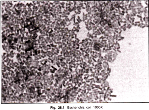
E. coli is Gram-negative straight rod, 1-3 µ x 0.4-0.7 µ, arranged singly or in pairs (Fig. 28.1). It is motile by peritrichous flagellae, though some strains are non-motile. Spores are not formed. Capsules and fimbriae are found in some strains.
3. Cultural Characteristics of Escherichia Coli:
It is an aerobe and a facultative anaerobe. The optimum growth temperature is 37°C. On Nutrient agar, colonies are large, thick, greyish white, moist, smooth, opaque or translucent discs. The smooth (s) form seen in fresh isolation is easily emulsified in saline, whereas the rough (R) form often auto agglutinates in saline.
Some strains may form “mucoid ” colonies. On MacConkey agar medium, colonies are bright pink due to lactose fermentation.
On selective media (Desoxycholate citrate agar-DCA; salmonella shigella-SS medium) used for the isolation of salmonella, their growth is inhibited, however their colonies are pink on DCA as it contains lactose and neutral red. In broth, there is generalized turbidity and deposit which disperses on shaking.
4. Biochemical Reaction of Escherichia Coli:
Glucose, lactose, mannitol, maltose are fermented with acid and gas production, but sucrose is not fermented by typical strain of E. coli. In Triple sugar iron (TSI), acid and gas are produced.
The four biochemical tests widely used for entero-bacteriaceae classification are Indole (I), Methyl Red (MR), Voges Proskauer (VP) and Citrate (C) utilisation which are referred to by the mnemonic IMV(1) C.E. coli is Indole and MR positive VP and citrate negative (IMV(1) C++ —), H2 S is not formed and urea is not hydrolysed.
5. Antigenic Structure of Escherichia Coli:
E. coli has Three Antigens:
O Somatic, Greek Ohne Hauch—without flagella; H—flagella; Greek Hauch—flagella and K (Kapsular) antigens. K antigen is an envelope antigen, which encloses the O antigen, renders the strain inagglutinable by the O antiserum and contributes to virulence by inhibiting phagocytosis.
It may be of three types —L, A and B. Though L type is common, the B antigen is medically important as it is found on enteropathogenic E. coli.
F (Fimbrial) Antigen:
The F antigen has no significance in antigenic classification of E. coli. Type I fimbriae mediates adhesion of bacterium to human and animal cells. Such adhesion enhances bacterial pathogenicity e.g. urinary tract infection in which type I fimbriae has some possible role to play.
Several fibrin structures resembling fimbriae have been demonstrated. They, most probably, play a very important role in pathogenesis of diarrhoeal diseases and urinary tract infection.
6. Toxin:
Besides the endotoxin associated with O antigen, some strains produce two types of exotoxin—enterotoxin and haemolysin. Enterotoxins responsible for diarrhoea are of two types—heat labile (LT); heat stable (ST).
LT is similar to cholera enterotoxin antigenically and in its mechanism of action—by stimulating the adenyl cyclase—cyclic adenosine monophosphate (cAMP) system to produce fluid accumulation in the intestinal lumen. ST appears to stimulate fluid secretion into the gut through the mediation of cyclic guanosine monophosphate (cGMP) resulting into dehydration.
7. Haemolysin by Escherichia Coli:
Three types of haemolysins produced by E. coli are not related to pathogenesis. E. coli forms a part of normal intestinal flora of man and animal and the commensal strains belong to several O groups. There are many strains of E. coli which include commensal strains as well as strains with virulence determinants that cause a wide variety of infections of all age groups of men and animals.
The virulent strains of E. coli are specific pathogens in the gut (enteritis) and of extra-intestinal sites (urinary tract infection, wound infection).
Clinical infections caused by Esch. coli are :
1. Urinary tract infection (UTI)
2. Septic infections of wound
3. Diarrhoea
4. Dysentery
5. Septicaemia
6. Pneumonia
7. Neonatal meningitis
8. Abscess in various organs.
Other pathogens of the family Entero-bacteriaceae causing UTI are Klebsiella, Proteus (P. mirabilis), Providence and Citrobacter. Gram-positive organisms (Staph, aureus, Enterococcus, Str. pyogenes) are also frequent pathogens of UTI.
The hospital acquired infections following instrumentation and catheterisation are mostly due to Pseudomonas and Proteus. Pregnant women (6-8%) do suffer from asymptomatic bacteriuria, if undetected and untreated, which may terminate into symptomatic infection later in pregnancy resulting to pyelonephritis and hypertension.
Calculi, enlarged prostate, pregnancy are predisposing factors in UTI. The reservoir of infection is the bacterial flora of colon. Urinary tract infection is usually from the perineum in ascending order via urethra.
A prerequisite for UTI is the colonisation of peri-urethral area by the pathogens. Due to shortness of urethra, the females are more prone to this infection. The haematogenous spread of this infection may be possible in newborn.
8. Infection: E. Coli Causes:
(1) Infantile acute diarrhoea or gastroenteritis;
(2) Urinary tract infection in pregnant women and men with prostatitis;
(3) Wound and burn infection, and
(4) Septicaemia.
1. Diarrhoea:
Four groups of E. coli responsible for diarrhoea in infants, children and adults are:
Enteropathogenic E. coli (EPEC); Enterotoxigenic E. coli (ETEC); Enteroinvasive E. coli (EIEC); Enterohaemorrhagic E. coli (EHEC).
EPEC:
EPEC is responsible for infantile diarrhoea. The pathogenic mechanism of EPEC has only recently been developed. They adhere to the intestinal mucosa, cause the loss of microvilli and prevent the entry of bacteria into the mucosa. They also produce a shigella-like toxin.
ETEC:
The enterotoxins are now known to produce diarrhoea in children with dehydration, traveller’s diarrhoea in adults, and sometimes cholera infantum similar to cholera. ETEC also possess colonisation factors (pili, K antigen) to enhance their virulence.
EIEC:
EIEC do not produce enterotoxin but invade the intestinal mucosa like dysentery bacilli. They cause kerato-conjunctivitis on instillation into the eyes of guinea pig (Sereny test) which is a diagnostic method for EIEC.
Another diagnostic method is their invasion of HeLa cells in tissue culture. EIEC is late lactose fermenter and may be anaerogenic. They have antigenic relationship with shigella.
EHEC:
EHEC has been very recently identified and is found to cause colitis with marked haemorrhage and absence of fever, produces Verotoxin (Cytotoxin) which affects the Vero cells in tissue culture.
9. Antigenic Typing of E. Coli:
Each E. coli has been primarily subdivided into a number of O groups which have been subdivided into subgroups with different K antigens. Each subgroup includes strains with different H antigens; separate numbers were previously allotted for B antigens, but afterwards these numbers have been included in the same consecutive series as the rest of K antigens.
Thus, a strain is recorded as 0111; K58; H12. The B number is no more in use, or, when used, the old number of B is written within brackets e.g. 0111; K58 (B4); H12.
a. Urinary tract infection:
It is caused by E. coli during catheterisation or pregnancy or due to urinary obstruction by prostitis. Pyelonephritis is produced due to haematogenous infection.
b. Pyogenic infection:
Superficial infections (wound or burn) or deep infections (peritonitis, cholecystitis, meningitis in children) are caused by E. coli.
c. Septicaemia:
E. coli, Pseudomonas aeruginosa or Gram-negative bacilli septicaemia is characterised by fever, hypotension, endotoxic shock (intravascular coagulation).
10. Laboratory Diagnosis of Escherichia Coli:
1. Diarrhoea:
For the detection of EPEC, fresh diarrhoeal stool is plated directly on blood agar and MacConkey agar medium. After overnight incubation, E. coli colonies are emulsified in saline on a slide and tested by agglutination with polyvalent and monovalent O antiserum against entero-pathogenic serotypes and further identified by biochemical tests.
Enzyme linked immuno-sorbent assay (ELISA) test is simplest and used to detect LT of ETEC. LT and cholera entero-toxin are antigenically similar. Sereney test is the only method available recently to demonstrate EIEC.
2. Urinary Tract Infection:
Catheterisation for urine collection is not advisable for diagnosis as it leads to urinary tract infection. Hence, in male, after cleaning the retracted prepuce and glans penis, with sterile cotton soaked in sterile normal saline, the first portion of urine that flushes out commensal bacteria from the anterior urether is discarded.
The next portion (midstream) of urine is collected in a sterile wide-mouthed container and despatched immediately to the laboratory as the urine is an ideal medium for the growth of bacteria. If there is a delay of 2-3 hours, it should be refrigerated.
Quantitative culture is done by pour plate or surface culture methods using ten-fold diluted urine. In the active urinary tract infection, the urine will contain 100,000 bacteria or more per ml. which is referred to as “significant bacteriuria,” counts of 10,000 bacteria or less are indicative of contamination.
The most commonly used “standard loop method” can transfer a fixed small volume of urine on non-inhibitory medium (Blood agar) for estimation of bacteria and on an indicative medium (MacConkey medium) for presumptive diagnosis of bacteriuria. The isolates are further identified and tested for antibiotic sensitivity. Antibiotic sensitivity may also be done directly using the urine samples.
Other screening techniques for the presumptive diagnosis of significant bacteriuria are:
(1) Griess nitrite test – The presence of nitrite indicates the presence of nitrate reducing bacteria in urine but nitrite is absent in normal urine.
(2) Catalase test – The frothing on addition of hydrogen peroxide due to catalase is an indication of bacteriuria.
3) Triphenyl tetrazolium chloride test is based on the production of pink red precipitate in the reagent due to the respiratory activity of growing bacteria.
(4) Microscopic demonstration of bacteria in Gram stained film of urine.
(5) Glucose test paper is based on the utilisation of minute amount of glucose present in normal urine by bacteria causing the infection.
(6) Dip slide culture method – Agar coated slides are immersed in the urine or exposed to the stream of urine are incubated and the colonies are estimated by colony counter. These screening tests are not sensitive and reliable as cultural test.
The antibody coated bacteria test has been employed for localisation of the site of urinary infection. Pyogenic infection and septicaemia diagnosis depends on the isolation of bacteria on culture.
11. Treatment of Escherichia Coli:
Infantile diarrhoea can be treated by Oral Rehydration Solution (ORS) as per World Health Organisation (WHO recommendation).
12. Medical Importance of Escherichia Coli:
E. coli is medically important because it
(a) Is used to produce insulin by adopting the genetic engineering technique,
(b) Produces certain vitamins in the intestine,
(c) Is used as parameter to determine the faecal contamination of drinking water,
(d) Is used for the plasmid study in the bacterial genetics.


